Amphibians, or amphibians, as adults are usually terrestrial animals, but they are still closely associated with the aquatic environment, and their larvae constantly live in water. Consequently, the Russian and Greek (amphibios - leading a double life) names reflect main feature these vertebrates. Amphibians originated, as mentioned above, from Devonian lobe-finned fish that lived in small fresh water bodies and crawled to the shore with the help of their fleshy paired fins.
External building. The body (Fig. 147) consists of the head, torso, front and rear paired dismembered limbs. The limbs consist of three sections: the front ones - from the shoulder, forearm and hand, the rear - from the thigh, lower leg and foot. Only a minority of modern amphibians have a tail (order caudates - newts, salamanders, etc.). It is reduced in adult forms of the largest group of amphibians - anurans (frogs, toads, etc.) due to the latter's adaptation to movement by jumping on land, but is preserved in their larvae - tadpoles living in water. In a few species leading a semi-subterranean lifestyle (the order legless, or caecilians), the limbs and tail were reduced.
The head is movably articulated with the body, although its movement is very limited and there is no pronounced neck. Dismembered limbs and a movable connection between the head and the body are characteristic features terrestrial vertebrates, they are absent in fish. The body of terrestrial forms is flattened in the dorso-ventral direction, while in fish (due to their adaptation to swimming) it is, as a rule, compressed laterally. In aquatic amphibians, the body shape approaches that of a fish. Body size ranges from 2 to 160 cm (Japanese salamander); On average, amphibians are smaller in size than other land animals. Skin bare rich in glands, separated in many places from the muscles due to the presence of subcutaneous lymphatic cavities. It is equipped with a large number blood vessels and also performs a respiratory function (see below). In some species, secretions from the skin glands are poisonous. Skin color is very diverse.
Nervous system. In connection with the adaptation of amphibians to life on land and especially in connection with the radical change in the nature of movement, the nervous system has changed quite a lot. Forebrain in amphibians (see Fig. 133, B) greater than average; in fish, as a rule, the opposite ratio is observed. This is explained by the fact that in fish the functions of the forebrain are associated only with the perception of olfactory stimuli; in amphibians, it begins to take part in the coordination of various functions of the body, and in its surface layer the rudiments of the cortex appear (still very weak), in which nerve cells. At the same time, it should be noted that the olfactory lobes are well developed in the forebrain. The cerebellum in amphibians is very poorly developed, unlike in fish. Fish are constantly moving, and their body position is unstable, while amphibians, leaning on their legs, are in a fairly stable position. The areas of the spinal cord where the nerves depart from it and go to the leg muscles, which perform much more work than the muscles of the paired fins of fish, are thickened and the brachial and lumbar plexuses of nerves are connected to them. The peripheral nervous system has changed greatly due to the differentiation of muscles (see below) and the appearance of long, jointed limbs.

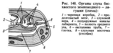
Of the sense organs, the organ of hearing has undergone the most significant changes. Transmission of sound waves from aquatic environment in animal tissue, which are also saturated with water and have approximately the same acoustic properties as water, occurs much better than from air. Sound waves, propagating in the air, are almost entirely reflected from the surface of the animal and only about 1% of the energy of these waves penetrates its body. In this regard, amphibians developed in addition to the labyrinth, or inner ear, a new section of the hearing organ is the middle ear. It is (Fig. 148) a small cavity filled with air, communicating with the oral cavity through the Eustachian tube and closed from the outside by a thin, elastic eardrum. In the middle ear there is an auditory plate (or column), which at one end rests against the eardrum, and at the other against a window covered with a film and leading into the cranial cavity, where there is a labyrinth surrounded by perilymph. The pressure inside the middle ear is equal to atmospheric pressure and the eardrum can vibrate under the influence of sound air waves, the impact of which is further transmitted through the auditory ossicle and perilymph to the walls of the labyrinth and is perceived by the endings of the auditory nerve. The cavity of the middle ear was formed from the first gill slit, and the column was formed from the hyomandibular bone (hyomandibular bone) located near the slit, which suspended the visceral part of the skull to the brain where the labyrinth was located behind the ear bones.
The eyes are covered with movable eyelids, which protect the organs of vision from drying out and clogging. Thanks to changes in the shape of the cornea and lens, amphibians see further than fish. Amphibians perceive small temperature changes well. They are sensitive to the effects of various substances dissolved in water. Their olfactory organ reacts to irritations caused by gaseous substances. Thus, the sensory organs of amphibians have undergone a number of changes in connection with the transition to living on land. Larvae and adult animals that live constantly in water have, like fish, lateral line organs.
Amphibians are characterized by rather complex instinctive actions, especially during the breeding season. For example, the male midwife toad, which lives in Russia in western Ukraine, wraps “cords” of eggs around its hind limbs and hides in secluded places on the shore until the tadpoles develop. After 17-18 days, the male returns to the water, where the tadpoles hatch. This is a kind of instinct to protect offspring. Even more complex instincts are known in a number of tropical tailless amphibians. Amphibians are also characterized by conditioned reflexes, however, they are produced with great difficulty.
Motor system and skeleton. The muscular system, in connection with various adaptations to life on land (the development of land-type limbs, the emergence of a movable joint between the head and the body, etc.) underwent radical transformations, although it retained many of the features inherent in fish. The muscular system of fish is very uniform and mainly consists of similar lateral muscle segments. In amphibians, the muscular system has become more differentiated, consisting of a variety of muscles (Fig. 149). Amphibians have the foundations of that muscular system, which then developed and became more complex in real land vertebrates - reptiles, birds and mammals. This also applies to the skeleton.
The skull of amphibians has many cartilaginous elements, which is probably explained by the need to lighten body weight due to a semi-terrestrial lifestyle. The skull contains many bones listed in the description of the skull of higher fish, including the parasphenoid characteristic only of fish and amphibians (Fig. 150). Since the hyomandibular bone has become an auditory bone, the role of the pendant is played by the quadrate bone. Due to the loss of the gill apparatus in adulthood, the gill arches are reduced and only their modified remains are preserved. The hyoid arch changes greatly and is partially reduced. The skull of amphibians is very wide, which is partly due to the characteristics of their breathing. Lower jaw, like bony fish, consists of several bones.
The vertebral column (Fig. 150) in tailless animals is very short and ends in a long bone - the urostyle, formed from the rudiments of the caudal vertebrae. In tailed amphibians, the caudal region spinal column consists of a number of vertebrae. In these amphibians, the tail plays a significant role in movement: in water it is used for swimming, on land it is used to maintain balance. The ribs are poorly developed (in caudate amphibians) or reduced, and their remains are fused with the transverse processes of the vertebrae (in other amphibians); ancient amphibians had ribs. Their reduction modern forms is explained by the need to lighten the body weight (which greatly increased during the transition from the aquatic environment to the air) in these vertebrates, which are not yet sufficiently adapted to movement on land. Due to reduction of ribs, amphibians do not have chest. The first vertebra is structured differently than in fish: it has two articular sockets for articulation with the two occipital condyles of the skull, due to which the head of amphibians has become mobile.
The skeleton of the forelimb (Fig. 150) consists of humerus, two bones of the forearm - the radius and the ulna, the bones of the wrist, the metacarpal bones and the phalanges of the fingers. The skeleton of the hind limb (Fig. 150) consists of the thigh, two bones of the lower leg - the tibia and fibula, tarsal bones, metatarsal bones and phalanges of the fingers. Consequently, the similarity in the structure of both pairs of limbs, despite some differences in their functions, is very great. Initially, the front and hind legs were five-toed; modern amphibians may have fewer toes. The hind limbs of many tailless amphibians are also used for swimming, and therefore they are elongated, and the fingers are connected by swimming membranes.
The limb girdles are much better developed than those of fish. The shoulder girdle consists of bone and cartilaginous elements: scapula, clavicle, crow bone (coracoid), etc. (Fig. 150). The clavicles and coracoids are connected to the sternum, which also includes bone and cartilaginous elements. The head of the humerus articulates with the shoulder girdle. The posterior girdle of the limbs, or pelvis, consists of three bones: the ilium, the pubis and the ischium (Fig. 150). The large acetabulum formed by these bones serves for articulation with the head of the femur. The pelvis is connected to one vertebra - the sacral one, thanks to which the hind legs, unlike the ventral fins of fish, received quite strong support.
Circulatory system. In the larvae of amphibians that live in water and breathe with gills, the circulatory system is basically similar to the circulatory system of fish, but in adult animals leading a terrestrial lifestyle, it changes significantly due to the replacement of gill respiration with pulmonary respiration, increased skin respiration, and the development of the limbs of land animals. type and other body changes. The heart (see Fig. 134, B, 151) consists of three chambers: the right and left atria and one ventricle. Departs from the right side of the latter conus arteriosus(it was also present in fish, the ancestors of amphibians), from which four pairs of arteries originate: the first pair - carotid arteries , carrying blood to the head, the second and third pairs are vessels connecting at the very large vessel bodies - aorta, the branches of which are directed to different parts of the body, the fourth pair - pulmonary arteries, which are then divided into independent cutaneous and pulmonary arteries.
From the lungs, oxygenated blood flows through the pulmonary veins into the left atrium, and blood, saturated in all parts of the body with carbon dioxide, flows into the anterior vena cava in the anterior part of the body, and into the posterior vena cava in the posterior part of the body (Fig. 152 ). Both vena cavae empty into venous sinus, from where blood (saturated with carbon dioxide) flows into the right atrium. From both atria, blood enters the single ventricle of the heart. The inner surface of the ventricle has depressions and therefore the blood in it does not have time to completely mix: in the left part there is blood saturated with oxygen, in the right part there is blood saturated with carbon dioxide, and in the middle part it is mixed. Since the conus arteriosus begins with right side ventricle, then the first portion of blood entering it (i.e., the arterial cone) will be venous, it is sent to the most posterior arteries - the pulmonary ones.
The mixed blood that then flows into the arteries that form the aorta, and through the branches of the latter into all parts of the body. Oxygenated blood from the left side of the ventricle is sent to the carotid arteries. To this it must be added that blood, saturated with oxygen in the skin, enters, as noted above, through the anterior vena cava and venous sinus into the right atrium and thus dilutes the venous blood located there, which is then pushed into the vessels that form the aorta. Consequently, thanks to the devices described above, as well as others not described here, different parts of the body receive blood unequally saturated with oxygen. In Fig. 152 shows the main arterial and venous vessels amphibians.
In amphibians, due to the strong development of the limbs and greater body dissection than in fish, the network of blood vessels has changed significantly. Many new vessels appeared that were absent in fish, and a system of vessels characteristic of terrestrial vertebrates emerged. At the same time, it should be remembered that the circulatory system of amphibians is much simpler than that of higher vertebrates.
Respiratory system. Almost all amphibians have lungs (see Fig. 151; 153). These organs still have a very simple structure and are thin-walled sacs, in the walls of which a rather dense network of blood vessels branches. Since the inner wall of the lungs is almost smooth, their surface area is relatively small. The trachea is almost undeveloped and the lungs are connected directly to the larynx. Since amphibians do not have a chest (see above), the act of breathing is ensured by the work of muscles oral cavity. Inhalation occurs as follows. With open nostrils (which, unlike the nostrils of fish, are through, i.e., in addition to the external nostrils there are also internal nostrils - choanae) and the mouth is closed, the bottom of the large oral cavity is pulled back and air enters it. Then the nostrils are closed with special valves, the bottom of the mouth is raised and air is forced into the lungs. Exhalation occurs as a result of contraction of the abdominal muscles.
Amphibians receive a significant amount of oxygen through the skin and mucous membranes of the oral cavity. Some species of salamanders have no lungs at all and all gas exchange occurs through the skin. However, the skin can only perform breathing functions if it is moist. Therefore, it is impossible for amphibians to live in conditions where air humidity is insufficient for them. Larvae living in water breathe through gills (first external, then internal) and skin. Some tailed amphibians that constantly live in water have gills that remain throughout their lives. Thus, in terms of breathing methods, amphibians are still close to fish.
Excretory system. The kidneys (see Fig. 136, A, B; Fig. 151), like those of fish, are trunk. Wolffian canals empty into the cloaca. The bladder opens there, where urine accumulates. Removal of dissimilation products also occurs through the skin and lungs.
Digestive system. The oral cavity is very wide. A number of species (mainly tailed amphibians) have many small, uniform, primitively arranged teeth that sit on the jaws, vomer, palatine and other bones and serve only to hold prey. In most species (mainly tailless amphibians), teeth are partially or completely reduced, but their tongue develops greatly. The latter in frogs is attached at the front end and can be thrown far forward with the rear end to catch prey. It is very sticky and well suited to perform specified function. In species that constantly live in water, the tongue is usually reduced. The capture of prey in such amphibians is carried out by the jaws.
The digestive tube (see Fig. 151) is relatively short and consists of the pharynx, esophagus, stomach, small intestine and a very small rectum (colon). The posterior part of the rectum is the cloaca; through it, in addition to feces, urine and sexual products are excreted. Salivary glands, which are absent in fish, flow into the oral cavity. The secretion of these glands serves mainly to moisten food. The salivary glands are very poorly developed in species that live in water, and much better in terrestrial ones. The liver is large; The pancreas is well defined. The food of adult amphibians is mainly animal (insects, small vertebrates, etc.). Tadpoles of tailless amphibians are mostly herbivorous.

Reproduction. The male gonads (testes) lie near the kidneys (see Fig. 151, B). Their ducts open into the tubules of the anterior part of the kidneys (see Fig. 136, A) and the seed is excreted, like urine, through the Wolffian canals. The female gonads (ovaries) grow greatly during the spawning period. The eggs exit through very long Müllerian canals (see Fig. 136, B). The latter do not have a direct connection with the ovaries and the ripening eggs enter through the body cavity into the funnels of the Müllerian canals.
Fertilization in most cases occurs in water. In many amphibians, this is preceded by the rapprochement of the male and female: the male clasps the female from behind, presses his forelimbs on her abdominal wall and this facilitates the release of eggs into the water, which he immediately fertilizes. Thus, in the presence of sexual intercourse, fertilization occurs outside the female’s body. In a minority of species (for example, newts), the male releases the seed in a special sac (spermatophore), which the female immediately captures with the edges of the cloaca. In this case, there is no sexual intercourse, but fertilization is internal. Finally, in some species the male inserts seed into the female's cloaca using his protruding cloaca.
In many species, sexual dimorphism is well expressed (in color, the structure of the front legs with which males hold females, and other characteristics). Males of a number of species can produce very loud sounds due to the amplification of these sounds by vocal sacs - resonators.
Development. Amphibian development usually occurs in water. From fertilized eggs, larvae (tadpoles) develop, which have a fish-like shape. They breathe through gills and their internal structure resembles fish. During the growth period, transformation (metamorphosis) of tadpoles occurs: first, their hind legs grow, then their front legs, gills and tail atrophy (in tailless animals), lungs develop, major changes occur in the circulatory system, etc.
Origin. Amphibians, as explained above (p. 296), descended from lobe-finned fish. The paired fins of ancient lobe-finned fish, from which the articulated limbs of terrestrial vertebrates developed, were short and wide, they included many small bone elements, not connected by joints, located in several (at least eight) transverse rows. The girdles on which the fins rested were relatively poorly developed (especially the pelvic girdle). Due to the transformation of fins into terrestrial limbs, significant changes occurred in the skeleton.
Firstly, many bone elements were reduced: in the first proximal rows there was only one bone left, in the front leg - the shoulder, in the back - the femur; in the second rows - two bones, in the front leg - radius and ulna, in the back - the tibia and fibula; in the next two rows, five bones remained, forming the carpus in the front leg and the tarsus in the back leg; in the next row, the remaining five bones were included in the metacarpus in the front leg, and the metatarsals in the rear leg; the remaining three rows with five bones each became the phalanges of the fingers. Reducing the number of bones contributed to increased strength of the legs.
Secondly, the bones of the first two rows (i.e., shoulder and forearm, thigh and lower leg) were greatly lengthened, which was very important for enhancing the speed of movement.
Thirdly, joints developed between the listed bones, i.e. the limbs became articulated, which is the most important condition their work.
Fourthly, the limb belts have been strengthened (see above for the description of the belts). Parallel to these changes, profound changes occurred in the nervous, muscular and vascular systems legs The changes in other organ systems that occurred during the transformation of lobe-finned fish into amphibians are described in general characteristics the latter.
The most ancient amphibians were stegocephalians (Fig. 154), which were numerous in the Carboniferous period and finally became extinct in the Triassic. They lived along the banks of reservoirs and spent a lot of time in the water. The head of these animals was covered with scutes, which explains their name (stegocephals - covered-headed). Their skeleton had many primitive features: the bone elements of the legs were small and slightly different in size, the vertebrae were biconcave, the girdles of the limbs were poorly developed, etc. Modern groups of amphibians originated from them.
Structure of the brain of bony fish
The brain of bony fishes consists of five sections typical for most vertebrates.
Diamond brain(rhombencephalon)
the anterior section extends under the cerebellum, and the posterior part, without visible boundaries, passes into spinal cord. To view the anterior part of the medulla oblongata, it is necessary to turn the body of the cerebellum forward (in some fish the cerebellum is small and the anterior part of the medulla oblongata is clearly visible). The roof of this part of the brain is represented by the choroid plexus. Underneath lies a large widened at the anterior end and passing behind into a narrow medial fissure, it is a cavity The medulla oblongata serves as the origin of most of the brain nerves, as well as a pathway connecting various centers of the anterior parts of the brain with the spinal cord. However, the layer of white matter covering medulla, in fish is quite thin, since the body and tail are largely autonomous - they carry out most of the movements reflexively, without relating to the brain. In the bottom of the medulla oblongata in fish and tailed amphibians lies a pair of giant Mauthner cells, associated with acoustic-lateral centers. Their thick axons extend along the entire spinal cord. Locomotion in fish is carried out mainly due to rhythmic bending of the body, which, apparently, is controlled mainly by local spinal reflexes. However, overall control over these movements is exercised by Mauthner cells. The respiratory center lies at the bottom of the medulla oblongata.
Looking at the brain from below, you can distinguish the origins of some nerves. Three round roots extend from the lateral side of the anterior part of the medulla oblongata. The first, lying most cranially, belongs to V and VII nerves, middle root - only VII nerve, and finally, the third root, lying caudally, is VIII nerve. Behind them, also from the lateral surface of the medulla oblongata, the IX and X pairs extend together in several roots. The remaining nerves are thin and are usually cut off during dissection.
Cerebellum Quite well developed, round or elongated, it lies over the anterior part of the medulla oblongata directly behind the optic lobes. With its posterior edge it covers the medulla oblongata. The part that protrudes upward is body of the cerebellum (corpus cerebelli). The cerebellum is the center for the precise regulation of all motor innervations associated with swimming and grasping food.
Midbrain(mesencephalon) - part of the brain stem penetrated by the cerebral aqueduct. It consists of large, longitudinally elongated optic lobes (they are visible from above).
Optic lobes, or visual roof (lobis opticus s. tectum opticus) - paired formations separated from each other by a deep longitudinal groove. The optic lobes are the primary visual centers for sensing stimulation. They're running out of fiber optic nerve. In fish, this part of the brain is of primary importance; it is the center that has the main influence on the activity of the body. The gray matter covering the optic lobes has a complex layered structure, reminiscent of the structure of the cerebellar cortex or hemispheres
Thick optic nerves arise from the ventral surface of the optic lobes and cross beneath the surface of the diencephalon.
If you open the optic lobes of the midbrain, you can see that in their cavity a fold is separated from the cerebellum, called cerebellar valve (valvule cerebellis). On either side of it in the bottom of the midbrain cavity there are two bean-shaped elevations called semilunar bodies (tori semicircularis) and being additional centers of the statoacoustic organ.
Forebrain(prosencephalon) less developed than the middle one, it consists of the telencephalon and diencephalon.
Parts diencephalon lie around a vertical slit Lateral walls of the ventricle - visual cusps or thalamus ( thalamus) in fish and amphibians are of secondary importance (as coordinating sensory and motor centers). The roof of the third cerebral ventricle - the epithalamus or epithalamus - does not contain neurons. It contains the anterior choroid plexus (choroid cover of the third ventricle) and the superior medullary gland - pineal gland (epiphisis). The bottom of the third cerebral ventricle - the hypothalamus or hypothalamus in fish forms paired swellings - lower lobes (lobus inferior). In front of them lies the inferior medullary gland - pituitary gland (hypophisis). In many fish, this gland fits tightly into a special recess in the bottom of the skull and usually breaks off during preparation; then clearly visible funnel (infundibulum). optic chiasm (chiasma nervorum opticorum).
in bony fishes it is very small compared to other parts of the brain. Most fish (except lungfishes and lobe-finned fish) are distinguished by the everted (inverted) structure of the telencephalon hemispheres. They seem to be “turned” ventro-laterally. The roof of the forebrain does not contain nerve cells and consists of a thin epithelial membrane (pallium), which during dissection is usually removed along with the membrane of the brain. In this case, the preparation shows the bottom of the first ventricle, divided into two by a deep longitudinal groove striatum. Striatum (corpora striatum1) consist of two sections, which can be seen when viewing the brain from the side. In fact, these massive structures contain striatal and cortical material of a rather complex structure.
Olfactory bulbs (bulbus olfactorius) adjacent to the anterior margin of the telencephalon. They go ahead olfactory nerves. In some fish (for example, cod), the olfactory bulbs are placed far forward, in which case they connect to the brain olfactory tracts.
Cranial nerves of fish.
In total, 10 pairs of nerves extend from the fish’s brain. Basically (both in name and in function) they correspond to the nerves of mammals.
Structure of the frog brain
Brain frogs, like other amphibians, are characterized by the following features compared to fish:
a) progressive development of the brain, expressed in the separation of the paired hemispheres by a longitudinal fissure and the development of the gray matter of the ancient cortex (archipallium) in the roof of the brain;
b) weak development of the cerebellum;
c) weak expression of the bends of the brain, due to which the intermediate and middle sections are clearly visible from above.

Diamond brain(rhombencephalon)
Medulla oblongata (myelencephalon, medulla oblongata) , into which the spinal cord passes cranially, it differs from the latter in its greater width and the departure from its lateral surfaces of the large roots of the posterior cranial nerves. On the dorsal surface of the medulla oblongata there is diamond-shaped fossa (fossa rhomboidea), accommodating fourth cerebral ventricle (ventriculus quartus). On top it is covered with a thin vascular cap, which is removed along with the meninges. The ventral fissure, a continuation of the ventral fissure of the spinal cord, runs along the ventral surface of the medulla oblongata. The medulla oblongata contains two pairs of cords (bundles of fibers): the lower pair, separated by the ventral fissure, are motor, the upper pair are sensory. The medulla oblongata contains the centers of the jaw and sublingual apparatus, the organ of hearing, as well as the digestive and respiratory systems.
Cerebellum located in front of the rhomboid fossa in the form of a high transverse ridge as an outgrowth of its anterior wall. The small size of the cerebellum is determined by the small and uniform mobility of amphibians - in fact, it consists of two small parts, closely connected with the acoustic centers of the medulla oblongata (these parts are preserved in mammals as fragments of the cerebellum (flocculi)). The body of the cerebellum - the center of coordination with other parts of the brain - is very poorly developed.
Midbrain(mesencephalon) when viewed from the dorsal side, it is represented by two typical optic lobes(lobus opticus s. tectum opticus) , having the appearance of paired ovoid elevations forming the upper and lateral parts of the midbrain. The roof of the optic lobes is formed by gray matter - several layers of nerve cells. The tectum in amphibians is the most significant part of the brain. The optic lobes contain cavities that are lateral branches cerebral (Sylvii) aqueduct (aquaeductus cerebri (Sylvii), connecting the fourth cerebral ventricle with the third.
The bottom of the midbrain is formed by thick bundles of nerve fibers - cerebral peduncles (cruri cerebri), connecting the forebrain with the medulla oblongata and spinal cord.
Forebrain(prosencephalon) consists of the diencephalon and telencephalon, lying sequentially.
visible from above as a rhombus, with sharp angles directed to the sides.
Parts of the diencephalon lie around a vertically located wide fissure third cerebral ventricle (ventriculus tertius). Lateral thickening of the walls of the ventricle - visual cusps or thalamus. In fish and amphibians, the thalamus is of secondary importance (as coordinating sensory and motor centers). The membranous roof of the third cerebral ventricle - the epithalamus or epithalamus - does not contain neurons. It contains the superior medullary gland - pineal gland (epiphisis). In amphibians, the pineal gland already serves as a gland, but has not yet lost the features of the parietal organ of vision. In front of the epiphysis, the diencephalon is covered with a membranous roof, which orally turns inward and passes into the anterior choroid plexus (choroid tectum of the third ventricle), and then into the endplate of the diencephalon. Inferiorly the ventricle narrows, forming pituitary funnel (infundibulum), the inferior medullary gland is attached to it caudoventrally - pituitary gland (hypophisis). In front, on the border between the bottom of the terminal and intermediate sections of the brain, there is chiasma nervorum opticorum). In amphibians, most of the fibers of the optic nerves are not retained in the diencephalon, but go further to the roof of the midbrain.
Telencephalon its length is almost equal to the length of all other parts of the brain. It consists of two parts: the olfactory brain and two hemispheres, separated from each other sagittal (arrow-shaped) fissure (fissura sagittalis).
Hemispheres of the telencephalon (haemispherium cerebri) occupy the posterior two-thirds of the telencephalon and hang over the anterior part of the diencephalon, partially covering it. There are cavities inside the hemispheres - lateral cerebral ventricles (ventriculi lateralis), caudally communicating with the third ventricle. In the gray matter of the cerebral hemispheres of amphibians, three areas can be distinguished: dorsomedially there is the old cortex or hippocampus (archipallium, s. hippocampus), laterally - ancient bark(paleopallium) and ventrolaterally - the basal ganglia, corresponding striata (corpora striata) mammals. The striatum and, to a lesser extent, the hippocampus are correlative centers, the latter associated with olfactory function. The ancient cortex is an exclusively olfactory analyzer. On the ventral surface of the hemispheres, grooves are noticeable, separating the striatum from the ancient cortex.
Olfactory brain (rhinencephalon) occupies the anterior part of the telencephalon and forms olfactory lobes (bulbs) (lobus olfactorius), soldered in the middle with each other. They are separated from the hemispheres laterally by the marginal fossa. The olfactory lobes anteriorly contain the olfactory nerves.
10 pairs extend from the frog's brain cranial nerves. Their formation, branching and zone of innervation are not fundamentally different from those in mammals
Bird brain.

Diamond brain(rhombencephalon) includes the medulla oblongata and cerebellum.
Medulla oblongata (myelencephalon, medulla oblongata) behind it directly passes into the spinal cord (medulla spinalis). Anteriorly, it wedges between the optic lobes of the midbrain. The medulla oblongata has a thick bottom, in which lie the nuclei of gray matter - the centers of many vital functions of the body (including equilibrium-auditory, somatic motor and autonomic). The gray matter in birds is covered with a thick layer of white, formed by nerve fibers connecting the brain to the spinal cord. In the dorsal part of the medulla oblongata there is diamond-shaped fossa (fossa rhomboidea), which is a cavity fourth cerebral ventricle (ventriculus quartus). The roof of the fourth cerebral ventricle is formed by a membranous vascular tegmentum; in birds it is completely covered by the posterior part of the cerebellum.
Cerebellum in birds it is large and is represented practically only worm (vermis), located above the medulla oblongata. The cortex (gray matter located superficially) has deep grooves that significantly increase its area. The cerebellar hemispheres are poorly developed. In birds, the sections of the cerebellum associated with muscle sense are well developed, while the sections responsible for the functional connection of the cerebellum with the cerebral cortex are practically absent (they develop only in mammals). The cavity is clearly visible in the longitudinal section cerebellar ventricle (ventriculus cerebelli), as well as alternation of white and gray matter, forming a characteristic pattern tree of life (arbor vitae).
Midbrain(mesencephalon) represented by two very large ones, shifted to the side visual lobes (lobus opticus s. tectum opticus). In all vertebrates, the size and development of the optic lobes is related to the size of the eyes. They are clearly visible from the side and from the ventral side, while from the dorsal side they are almost completely covered by the posterior sections of the hemispheres. In birds, almost all the fibers of the optic nerve come to the optic lobes, and the optic lobes remain extremely important parts of the brain (however, in birds, the cerebral cortex begins to compete with the optic lobes in importance). The sagittal section shows that in the forward direction the cavity of the fourth ventricle, narrowing, passes into the cavity of the midbrain - cerebral or Sylvian aqueduct (aquaeductus cerebri). Orally, the aqueduct passes, expanding, into the cavity of the third cerebral ventricle of the diencephalon. The conventional anterior border of the midbrain is formed posterior commissure (comissura posterior), clearly visible on a sagittal section in the form of a white spot.
Included forebrain(prosencephalon) there are the diencephalon and the telencephalon.
Diencephalon in birds it is visible from the outside only from the ventral side. The middle part of the longitudinal section of the diencephalon is occupied by a narrow vertical fissure third ventricle (ventriculus tertius). In the upper part of the ventricular cavity there is a hole (paired) leading into the cavity of the lateral ventricle - Monroe (interventricular) foramen (foramen interventriculare).
The lateral walls of the third cerebral ventricle are formed by a fairly well developed thalamus, the degree of development of the thalamus is related to the degree of development of the hemispheres. Although it does not have the significance of a higher visual center in birds, it nevertheless performs important functions as a motor correlative center.
In the anterior wall of the third ventricle lies anterior commissure (comissura anterior), consisting of white fibers connecting the two hemispheres
The floor of the diencephalon is called hypothalamus (hypothalamus). When viewed from below, lateral thickenings of the bottom are visible - visual tracts (tractus opticus). Between them the anterior end of the diencephalon includes optic nerves (nervus opticus), forming optic chiasm (chiasma opticum). The posterior lower corner of the third cerebral ventricle corresponds to the cavity funnels (infunbulum). From below, the funnel is usually covered by the subcerebral gland, which is well developed in birds - pituitary gland (hypophysis).
From the roof of the diencephalon (epithalamus) extending upward having a cavity pedicle of the pineal organ. Above is himself pineal organ- pineal gland (epiphysis), it is visible from above, between the posterior edge of the cerebral hemispheres and the cerebellum. The anterior part of the roof of the diencephalon is formed by the choroid plexus extending into the cavity of the third ventricle.
Telencephalon in birds it consists of cerebral hemispheres (hemispherium cerebri), separated from each other by deep longitudinal fissure (fissura interhemispherica). The hemispheres in birds are the largest formations of the brain, but their structure is fundamentally different from that of mammals. Unlike the brain of many mammals, the greatly enlarged hemispheres of the bird's brain do not bear grooves and convolutions; their surface is smooth on both the ventral and dorsal sides. The cortex as a whole is poorly developed, primarily due to the reduction of the olfactory organ. The thin medial wall of the forebrain hemisphere in the upper part is represented by nerve substance old bark (archipallium). Material neocortex(poorly developed) (neopallium) along with a significant mass striatum (corpus striatum) forms a thick lateral wall of the hemisphere or a lateral outgrowth, protruding into the cavity of the lateral ventricle. Therefore the cavity lateral ventricle (ventriculus lateralis) hemisphere is a narrow gap located dorsomedially. In birds, unlike mammals, significant development in the hemispheres is achieved not by the cerebral cortex, but by the striatum. It has been revealed that the striatum is responsible for innate stereotypical behavioral reactions, while the neocortex provides the ability for individual learning. Some bird species have been found to have better-than-average development of a portion of the neocortex, such as crows, known for their learning abilities.
Olfactory bulbs (bulbis olfactorius) located on the ventral side of the forebrain. They are small in size and approximately triangular in shape. They enter from the front olfactory nerve.
137. Look at the pictures. Write the names of the frog's body parts. What organs are located on her head? Write their names.
138. Study the table “Class Amphibians. The structure of a frog." Look at the drawing. Write the names internal organs frogs indicated by numbers.

139. Describe the structure of the amphibian brain.
The brain of amphibians has more progressive features: larger forebrain sizes, complete separation of the hemispheres. The midbrain is relatively small. The cerebellum is small because amphibians have monotonous movements. There are 10 pairs of cranial nerves leaving the brain. Divisions of the brain: anterior, middle, cerebellum, medulla oblongata, intermediate.
140. Study the table “Class Amphibians. The structure of a frog." Look at the drawing. Label the parts of the frog's skeleton indicated by numbers.

1. skull
2. shoulder blade
3. shoulder
4. forearm
5. brush
6. foot
7. shin
8. thigh
9. urostyle
10. spine.
141. Look at the drawing. Write the names of the parts of the frog's digestive system indicated by numbers. How is digestion carried out in a frog?

All amphibians feed only on mobile prey. At the bottom of the oral cavity is the tongue. When catching insects, it is thrown out of the mouth, and the prey sticks to it. On upper jaw There are teeth that serve only to hold prey. When swallowing, the eyeballs help push food into the esophagus from the oropharynx.
The ducts of the salivary glands open into the oropharynx, the secretion of which does not contain digestive enzymes. From the oropharynx cavity, food enters the stomach through the esophagus, from there to duodenum. The ducts of the liver and pancreas open here. Digestion of food occurs in the stomach and duodenum. Small intestine passes into the rectum, which forms an extension - the cloaca.
142. Draw a diagram of the structure of a frog’s heart. Which blood is called arterial and which is called venous?
Arterial blood comes from the lungs and is rich in oxygen. Venous blood goes to the lungs.

143. Describe the process of reproduction and development of a frog. Indicate the similarities in the reproduction of amphibians and fish.
Amphibians breed in shallow, well-warmed areas of water bodies. The reproductive organs of males are the testes, and the reproductive organs of females are the ovaries. Fertilization is external.
Frog development:
1 - egg;
2 - tadpole at the moment of hatching;
3 - development of fin folds and external gills;
4 - stage of maximum development of external gills;
5 - stage of disappearance of external gills; 6 - stage of appearance of the hind limbs; 7 - stage of dismemberment and mobility of the hind limbs (the forelimbs are visible through the integument);
8 - stage of release of the forelimbs, metamorphosis of the oral apparatus and the beginning of tail resorption;
9 - stage of landfall.
144. Fill out the table.
The structure and significance of the frog's sense organs.

145. Execute laboratory work"Peculiarities external structure frogs in connection with their way of life.”
1. Consider the features of the external structure of the frog. Describe the shape of its body, the color of its back and abdomen.
The frog's body is divided into head, torso and limbs. Long hind legs with webbed toes allow it to jump on land and swim in water. On the flattened head of the frog there is a large mouth slit, large bulging eyes located on elevations and a pair of nostrils. On the sides of the head behind the eyes are rounded eardrums (eardrums). The frog's eyes are large and bulging. The eyes are equipped with movable eyelids. Male green frogs have resonators, or vocal sacs, in the corners of their mouths, which inflate when they croak, amplifying the sounds.
The skin of amphibians is bare and moist, covered with mucus.
Body coloring helps protect against enemies.
2. Draw a drawing of the frog’s body and label its sections.

3. Consider the structure of the front and hind limbs. Sketch them.

4. Examine the frog's head. What sense organs are located on it?
see table No. 144
5. Note the structural features of the frog associated with life in water and on land.
In water: skin is bare, covered with mucus. There are nostrils on the head and eyes on the top. There are swimming membranes on the paws. Hind legs long. Development and reproduction in water. In water it switches to skin respiration. Cold-blooded. The larva has structural features similar to fish.
On land: 2 pairs of limbs, moves. Breathes with lungs. Feeds on insects. The heart is three-chambered.
Conclusions: amphibians are the first chordates to reach land. They still have external and internal structural features that allow them to partially live in water, however, they also have progressive structural features characteristic of terrestrial animals.
Muscular system. It differs from such fish mainly in the greater development of the muscles of the limbs and the greater differentiation of the trunk muscles, consisting of complex system individual muscles. As a result, the primary segmentation of the muscles is disrupted, although in some abdominal and dorsal muscles it still remains quite distinct.
Nervous system. The brain of amphibians differs from the brain of fish mainly in the greater development of the forebrain, the complete separation of its hemispheres and the underdeveloped cerebellum, which is only a small ridge of nerve substance covering the anterior part of the fourth ventricle. The development of the forebrain is expressed not only in its enlargement and differentiation, but also in the fact that, in addition to the bottom of the lateral ventricles, their sides and roof contain nervous substance, i.e. in amphibians a real brain vault appears - the archipallium (from modern fish the archipallium is present in lungfishes). The olfactory lobes are only weakly delimited from the hemispheres. The diencephalon is only slightly covered from above by neighboring sections. The parietal organ is attached to its roof, and a well-defined funnel extends from the bottom, to which the pituitary gland is attached. Although the midbrain is a significant section, it is relatively smaller than that of fish. Underdevelopment of the cerebellum, as in lungfishes, is associated with the simplicity of body movements: amphibians are generally sedentary animals, but in those that, like frogs, can make rapid movements, they are limited to jumping, i.e., very simple movements. From the brain, like in bony fishes, only 10 pairs of head nerves depart; The XII pair (hypoglossal nerve) extends outside the cranium, and the XI pair (accessory nerve) is not developed at all.

. I - top; II - bottom; III - side; IV - in longitudinal section (according to Parker):
1 - forebrain hemispheres, 2 - olfactory lobe, 3 - olfactory nerve, 4 - diencephalon, 5 - optic chiasm, 6 -funnel, 7 - pituitary gland, 8 - midbrain, 9 - cerebellum, 10 - medulla oblongata, 11 - fourth ventricle, 12 - spinal cord, 13 - third ventricle, 14 - aqueduct of Sylvius,
III - X - head nerves, XII - hypoglossal nerve

, scheme (according to Gregory):
1 — skull, 2 - medulla oblongata, 3 - auditory nerve, 4 - semicircular canals, 5 - middle ear cavity, 6 - Eustachian tube, 7 - pharynx, 8 - three, 9 - eardrum
The frog has 10 pairs of true spinal nerves. The three front pairs take part in the formation brachial plexus, innervating the forelimbs, and the four rear pairs - in the formation of the lumbosacral plexus, innervating the hind limbs.
The sympathetic nervous system of the frog, like all amphibians, is very well developed and is represented mainly by two nerve trunks that stretch on both sides of the spine and are formed by a chain nerve ganglia, connected to each other by cords and connected to the spinal nerves.
More interesting articles
The frog is a typical representative of amphibians. Using this animal as an example, you can study the characteristics of the entire class. This article describes in detail the internal structure of a frog.
Begins digestive system oropharyngeal cavity. At its bottom is attached a tongue, which the frog uses to catch insects. Thanks to its unusual structure, it is capable of being thrown out of its mouth at high speed and sticking its victim to itself.
On the palatine bones, as well as on the lower and upper jaws of the amphibian, there are small conical teeth. They do not serve for chewing, but primarily for holding prey in the mouth. This is another similarity between the amphibian and fish. The secretion secreted by the salivary glands moistens the oropharyngeal cavity and food. This makes it easier to swallow. Digestive enzymes frog saliva does not contain.

The frog's digestive tract begins with the pharynx. Next comes the esophagus, and then the stomach. Behind the stomach is the duodenum, the rest of the intestine is laid out in the form of loops. The intestine ends in the cloaca. Frogs also have digestive glands - the liver and pancreas.
The prey caught with the help of the tongue ends up in the oropharynx, and then through the pharynx enters the esophagus into the stomach. Cells located on the walls of the stomach secrete hydrochloric acid and pepsin, which help digest food. Next, the semi-digested mass follows into the duodenum, into which the secretions of the pancreas also pour out and flow into bile duct liver.
Gradually the duodenum turns into small intestine, where everything is absorbed useful material. The remains of food that has not been digested end up in the last section of the intestine - the short and wide rectum, ending in the cloaca.
The internal structure of the frog and its larvae are different. Adults are predators and feed mainly on insects, but tadpoles are true herbivores. On their jaws there are horny plates, with the help of which the larvae scrape off small algae along with the single-celled organisms living in them.
Respiratory system
Interesting features of the internal structure of the frog also concern breathing. The fact is that, along with the lungs, the capillary-filled skin of the amphibian plays a huge role in the process of gas exchange. The lungs are thin-walled paired bags with a cellular inner surface and an extensive network of blood vessels.

How does a frog breathe? The amphibian uses valves capable of opening and closing its nostrils and movements of the floor of the oropharynx. In order to inhale, the nostrils open, and the bottom of the oropharyngeal cavity drops, and the air ends up in the frog's mouth. To allow it to pass into the lungs, the nostrils close and the floor of the oropharynx rises. Exhalation occurs due to the collapse of the pulmonary walls and movements of the abdominal muscles.
In males, the laryngeal cleft is surrounded by special arytenoid cartilages, on which the vocal cords are stretched. High sound volume is ensured by the vocal sacs, which are formed by the mucous membrane of the oropharynx.
Excretory system
The internal structure of the frog, or rather, it is also very curious, since the waste products of the amphibian can be excreted through the lungs and skin. But still, most of them are secreted by the kidneys, which are located at the sacral vertebra. The kidneys themselves are oblong bodies adjacent to the back. These organs have special glomeruli that are capable of filtering waste products from the blood.
Urine is discharged through the ureters into the bladder, where it accumulates. After filling Bladder the muscles at the abdominal surface of the cloaca contract and fluid is thrown out through the cloaca.
Circulatory system
The internal structure of the frog is more complex than that of an adult frog; it is three-chambered, consisting of a ventricle and two atria. Due to the single ventricle, arterial and venous blood are partially mixed, the two circulation circles are not completely separated. The conus arteriosus, which has a longitudinal spiral valve, extends from the ventricle and distributes mixed and arterial blood into different vessels.

Mixed blood collects in the right atrium: venous blood comes from the internal organs, and arterial blood comes from the skin. Arterial blood enters the left atrium from the lungs.
The atria contract simultaneously, and blood from both enters a single ventricle. Due to the structure of the longitudinal valve, it enters the organs of the head and brain, mixed - to organs and parts of the body, and venous - to the skin and lungs. Students may have a hard time understanding the internal structure of a frog. Scheme circulatory system amphibians will help you visualize how blood circulation works.
The circulatory system of tadpoles has only one circulation, one atrium and one ventricle, like in fish.
The structure of the blood of a frog and a person is different. have a core, oval shape, and in humans they have a biconcave shape, with no core.
Endocrine system
IN endocrine system frogs include the thyroid, reproductive and pancreas glands, adrenal glands and pituitary gland. Thyroid produces hormones necessary to complete metamorphosis and maintain metabolism; the gonads are responsible for reproduction. The pancreas is involved in the digestion of food, the adrenal glands help regulate metabolism. The pituitary gland produces a number of hormones that affect the development, growth and coloring of the animal.

Nervous system
The nervous system of the frog is characterized by a low degree of development; it is similar in characteristics to the nervous system of fish, but has more progressive features. The brain is divided into 5 sections: midbrain, diencephalon, forebrain, medulla oblongata and cerebellum. The forebrain is well developed and is divided into two hemispheres, each of which has a lateral ventricle - a special cavity.
Due to monotonous movements and a generally sedentary lifestyle, the cerebellum is small in size. The medulla oblongata is larger. In total, ten pairs of nerves emerge from the frog's brain.
Sense organs
Significant changes in the sensory organs of amphibians are associated with the exit from the aquatic environment to land. They are already more complex than those of fish, since they must help navigate both in water and on land. Tadpoles have developed lateral line organs.

Pain, tactile and temperature receptors are hidden in the epidermis layer. Papillae on the tongue, palate and jaws serve as taste organs. The olfactory organs consist of paired olfactory sacs, which open with both external and internal nostrils, in environment and the oropharyngeal cavity, respectively. In water, the nostrils are closed, the sense of smell does not function.
As a hearing organ, the middle ear is developed, in which there is an apparatus that amplifies sound vibrations thanks to the eardrum.
The structure of a frog's eye is complex, because it needs to see both underwater and on land. The eyes of adults are protected by movable eyelids and a nictitating membrane. Tadpoles do not have eyelids. The cornea of a frog's eye is convex, the lens is biconvex. Amphibians can see quite far and have color vision.


