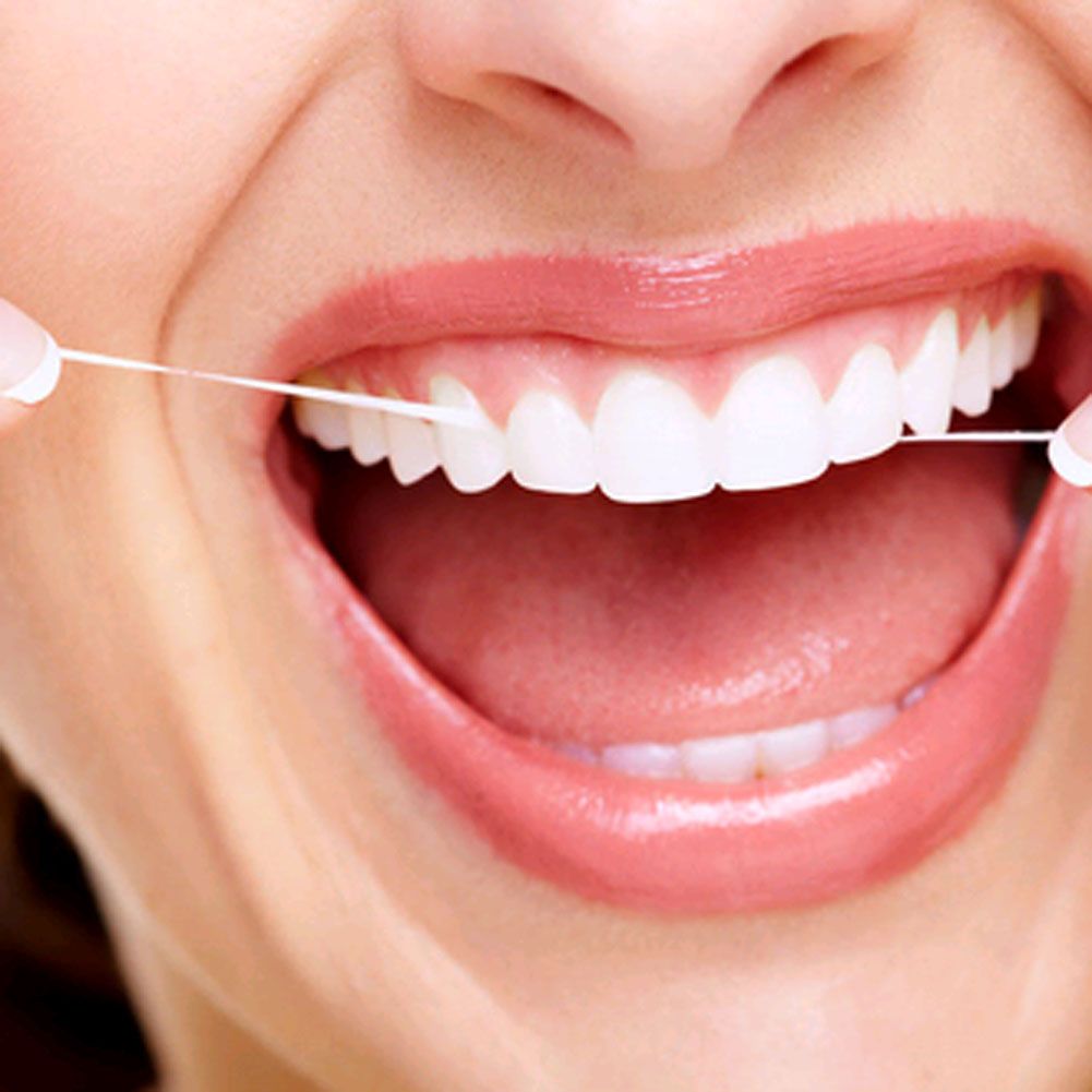
IN oral cavity the food is crushed, moistened with saliva, and then moved into the pharynx.
The organs of the oral cavity are: lips, cheeks, teeth, gums, hard and soft palate, tongue, tonsils and salivary glands. Its bone base is formed by the maxillary, mandibular, incisive and palatine bones. In the oral cavity, the vestibule and the oral cavity proper are distinguished. These parts are separated by teeth, incisor bones and gums. The vestibule of the mouth is in the form of a gap, bounded externally by the upper and lower lips and cheeks, and internally by two rows of teeth. The edges of the lips limit the entrance opening - the oral fissure. Actually, the oral cavity is limited in front and on the sides by the gums and teeth, above and behind by the hard and soft palate, and below by the bottom of the oral cavity with the tongue.
Lips consist of skin, muscle layer and mucous membrane.
The skin covers the outer surface of the lips. Under the skin lies a closely fused neuromuscular layer. Inner layer lips - mucous membrane. In the submucosa connective tissue labial glands are congested. The mucous membrane of the lips, like the entire oral cavity, is covered with squamous stratified epithelium. On the free edge of the lips it passes into the general skin. The lower lip continues into the chin.
U horses, sheep And goats the lips are strongly developed and easily mobile in all areas. In the middle, the labial groove is clearly visible (less marked in the horse).
Lips cattle fat and inactive. The upper lip passes into the nasolabial mirror, devoid of hairline. The mucous membrane of the lateral parts of the lips forms high cone-shaped papillae.
Lips pigs short and sedentary. The upper lip merges with the proboscis, the lower one becomes sharper.
U dogs the upper lip has a deep narrow groove and passes into the nasal mirror.
The cheeks form the side walls of the oral cavity. They consist of skin, muscle, glandular layer and mucous membrane. The buccal glands are divided into superior and inferior. Cattle also have middle buccal glands. The mucous membrane of the cheeks forms cone-shaped papillae.
The gums are a mucous membrane that covers the alveolar processes of the jaws and incisive bones, surrounds the necks of the teeth and closely fuses with the periosteum. In ruminants, in place of the upper incisor teeth, the gum forms a thickening - a dental plate, covered with a thick keratinizing epithelium.
Solid sky serves as the roof of the oral cavity and is formed by a thick, tough mucous membrane covering the bony palate. Aborally it passes into the soft palate, or velum, and laterally into the gums. A palatal suture runs along the midline of the hard palate, on both sides of which on the hard palate there are transverse thickenings of the mucous membrane - palatine ridges - located in the form of arched folds. At the anterior end of the palatal suture, directly behind the incisive teeth, the palatine, or incisive, papilla rises (the horse does not have one), on the sides of which thin nasolacrimal canals open into the nasal cavity.
In the submucosal tissue of the hard palate there is a dense plexus of venous vessels.
Soft sky, or velum, is a continuation of the hard palate. It is a fold of the mucous membrane with muscles, glands, and lymph nodes embedded in it and separates the oral cavity from the pharyngeal cavity. Between the free edge of the velum and the root of the tongue, a hole is formed leading from the oral cavity to the pharyngeal cavity. This is the pharynx slit. U horses The velum palatine is very long, reaching the root of the tongue, which eliminates the possibility of breathing through the mouth. Other species have shorter velum palates and can breathe through their mouths.
The velum palatine has two surfaces: one faces the pharynx, its mucous membrane is lined with a prismatic ciliated epithelium; the other is directed towards the mouth, its mucous membrane is lined with flat multilayered epithelium. The free, concave edge of the velum palatine is called the palatine arch, which passes into the velopharyngeal arches, running along the lateral walls of the pharynx to the dorsal wall of the esophagus and forming an unpaired esophagopharyngeal arch above the entrance to the esophagus.
The basis of the velum palatine is the muscular elephant, consisting of several muscles. The muscles carry out the movement of the palatine curtain. As a result of the contraction of these muscles, the velum palatine is shortened after the act of swallowing, rises during the act of swallowing and tenses, helping the tongue to form and push the food bolus into the pharynx.
Rice. 1. Language:
L - horses; B - cattle; D- sheep; G - pigs; D - dogs; 1 - roller-shaped papillae; 2 - leaf-shaped papillae; 3 -fungiform papillae; 4 - tongue pillow; 5 - tip of the tongue; 6 - body of the tongue; 7 - root of the tongue.
Tonsils. Between the velum palatine and the root of the tongue on the right and left lie the palatine tonsils with lymphatic follicles. In horses, on the pharyngeal and oral surfaces of the velum palatine, the mucous membrane contains lymphatic follicles that form the unpaired palatine tonsil. The tonsils function as the first protective devices in the fight against infection that enters the body through the mouth and nasal openings.
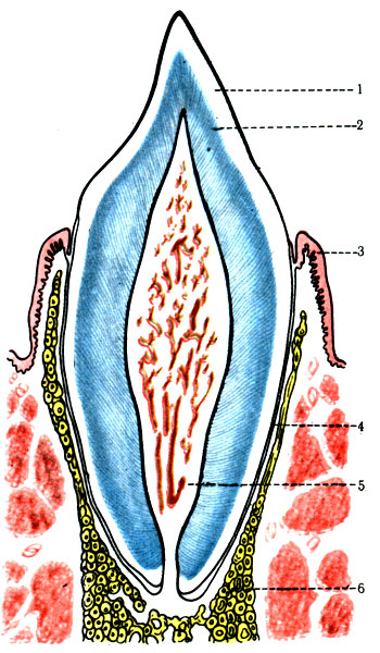
(Fig. 1) is a mobile muscular organ whose role is to capture food, place it on the teeth during chewing, move it from the oral cavity to the pharyngeal cavity, and determine its nature and quality. Language is divided into root, body and apex.
The root of the tongue extends from the larynx to the last molar, the body of the tongue lies between the molars, the apex of the tongue is the anterior free-lying part. The upper surface of the tongue is called its dorsum. The mucous membrane of the floor of the oral cavity passes to the lower surface of the tongue, forming a fold - the frenulum of the tongue. The mucous membrane of the upper and lateral surfaces of the tongue forms special protrusions - papillae: filiform, cone-shaped, ridge-shaped, mushroom-shaped, leaf-shaped. Filiform and cone-shaped papillae cover the back and tip of the tongue and have a mechanical significance - they help move food. Fungiform papillae are located mainly on the lateral surfaces of the body of the tongue and the upper surface of the tip of the tongue. The roller-shaped papillae are located on the back of the tongue; unlike the fungiform papillae, they are embedded in the thickness of the mucous membrane. Leaf-shaped papillae in the form of several parallel folds separated by narrow grooves are visible, one on each side, along the edges of the root of the tongue.
Serous glands open to the bottom of the grooves. The papillae of the tongue, with the exception of the filiform ones, contain taste buds and are an organ of taste. In the thickness of the mucous membranes of the root and lateral edges of the tongue there are many lymphatic follicles and mucous glands that secrete a secret that moisturizes the tongue.
The muscles of the tongue have longitudinal, transverse and vertical fiber directions. By contracting, they shorten, thicken, and narrow the tongue. The muscles coming from the hyoid bone and chin pull the tongue back, push it forward, carry out lateral movements, and push the food lump.
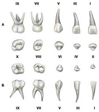
cattle hard, thick, on the back half of the back with an elevation - a pillow of the tongue. The filiform papillae are thick and large. The epithelium of the papillae is highly keratinized. There are from 8 to 17 roller-shaped papillae on each side of the tongue. There are no leaf-shaped papillae. pigs slightly pointed, relatively long and narrow. The filiform papillae are thin and soft. The fungiform papillae are small; there are only two valicular papillae, one on each side of the root of the tongue. horses long, tapering anteriorly. The filiform papillae are thin, long, soft, and create a velvety surface on the back of the tongue. There are two valicular papillae. Leaf-shaped papillae are elongated. dogs wide, flat, thin, with sharp lateral edges. There is a shallow groove on the back (in the middle). There are two valicular papillae.Teeth(Fig. 2). The function of teeth is to mechanically process food. In each tooth it is customary to distinguish a crown - a part that freely protrudes into the oral cavity; the neck, covered by the gum, and the root, immersed in the alveolus of the corresponding bone: the upper, lower jaw or incisor bones.
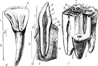
Rice. 2. Cow teeth:
A-incisor; B- longitudinal cut of the incisor tooth; B-structure of a molar; 1 -crown; 2 - root; h- neck; 4 - chewing surface; 5 - cement; 6 - enamel; 7 - dentin; 8 - tooth cavity, filled with pulp; O- cups; 10 - gums; 11 - mandibular bone with dental alveolus; 12 - periosteum of the socket.
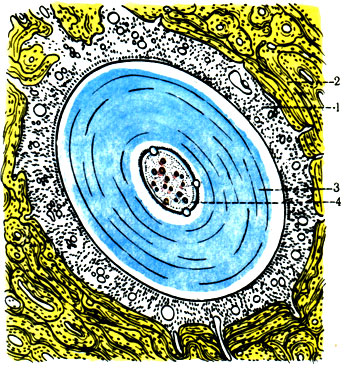
Each tooth consists of dentin, enamel, cementum and pulp. The main part is dentin. Inside the tooth, starting from the end of the root, there is dental cavity, filled with dental pulp, in which blood vessels and nerves branch.
There are short-crowned and long-crowned teeth. In short-crowned teeth, enamel covers dentin only in the crown area, in the form of a cap, and cement covers the root of the tooth. In long coronal teeth, not only the crown, but also the root of the tooth is covered with enamel, and the entire tooth on top of the enamel and even the cups formed by folds of enamel on the rubbing surface of the tooth are covered with cement. The crown of the tooth is very long and moves out of the dental socket as it wears away. Short-crowned teeth include all milk teeth, permanent incisors of cattle, all teeth of pigs and dogs; to long-crowned - all the permanent teeth of a horse and the permanent molars of cattle.
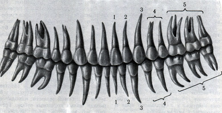
The teeth are arranged in the form of dental arcades and are divided into incisors, canines and molars. Domestic animals have six incisors on their lower jaws and incisive bones. The exception is ruminants, which have eight incisors on the lower jaws and no teeth on the incisal bones.
The incisors have the following names (counting from the middle): hooks, middle and edges; in ruminants, the lower jaws have middle medial (next to the toes) and middle lateral (next to the margins). The incisors grab the food and chew it off.
The canines are located one on each side in the upper and lower arcades. Ruminants do not have fangs. Fangs serve as weapons of defense and attack.
Horses and ruminants have six molars on each jaw, pigs have seven, and dogs have seven (on the lower jaws). The food is ground with molars. Three or four front molars on each side have milk predecessors - premolars. The posterior molars, molars, and milk predecessors do not have.
U ruminants 20 primary teeth (8 incisors on the lower jaws and 12 premolars) and 32 permanent teeth(8 incisors, 12 premolars and 12 molars). The molars of ruminants are of the lunate type.
U pigs 28 primary teeth (12 incisors, 4 canines and 12 premolars) and 44 permanent teeth (12 incisors, 4 canines, 16 premolars and 12 molars). The permanent molars of pigs are of the tuberculate type.
U dogs 32 primary teeth (12 incisors, 4 canines and 16 molars) and 42 permanent teeth (12 incisors, 4 canines, 26 molars, including 16 premolars and 10 molars). The permanent teeth of dogs are three-toothed and multi-cusped.
U horses 24 baby teeth (12 incisors and 12 premolars) and 40 permanent teeth in males (12 incisors, 4 canines, 12 premolars and 12 molars). Mares have rudimentary canines and often do not have them. Incisive teeth are in the shape of a curved wedge with a convex, labial, and concave, lingual, surface.
The structure of the teeth is in accordance with the chewing nature of animals. In herbivores, this is accomplished through grinding movements of the lower jaw down and up, but also sideways and forward. The chewing surfaces of the molars are located like millstones, one above the other and grind everything that is between them. Ruminants grind food especially carefully when they regurgitate the cud.
In pigs and dogs, the lower jaw can only be lowered and raised, which achieves cutting and crushing of feed masses.
Determining the age of domestic animals by teeth is based on the time of appearance of milk teeth and their replacement by permanent ones, and in horses, in addition, on the abrasion and change in the shape of the rubbing surface of the incisor teeth.
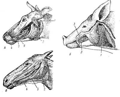
Rice. 3. Parietal and parietal salivary glands:
A- cattle; B- pigs; IN- horses; 1 - parotid salivary gland; 2 - submandibular salivary gland; h- buccal glands; 4 - labial glands; 5 - duct of the submandibular salivary gland; 6 - sublingual short-duct salivary gland; 7 - sublingual salivary gland ductal.
The replacement of primary incisors with permanent ones begins in foals at 2.5 years of age and ends by 5 years of age. Primary incisors are easy to distinguish from permanent incisors: they are whiter, smaller in size, with a clearly defined neck and a shallow (up to 4 mm) cup. In young animals, the upper and lower incisor teeth are located towards each other in the form of an arc. The angle at which the incisor teeth meet becomes sharper as the animal ages.
Form cross section the rubbing surface of the permanent incisors changes with age in the following order: in foals it is transversely oval, in middle-aged horses it is round; in old animals it is triangular, in very old animals it is obverse. During this period, the dental cup disappears, and a cup mark and dental star appear.
Salivary glands(Fig. 3). In the wall of the mucous membrane of the lips, cheeks, tongue, and palate, the parietal salivary glands are embedded in the form of separate formations or groups. Outside the oral cavity there are large parotid salivary glands: paired parotid, sublingual and submandibular. The secretion of the salivary glands, which flows into the oral cavity through the excretory ducts, is called saliva.
Functionally, the salivary glands are divided into serous, mucous and mixed. The secretion of the serous glands contains a lot of protein, which is why they are also called proteinaceous. The secretion of the mucous glands contains the mucous substance mucin. Mixed glands secrete a protein-mucus secretion.
The parotid salivary gland is serous (mixed in some areas in carnivores), and alveolar in structure. In cattle, pigs and dogs it is triangular in shape, in horses it is rectangular. Lies approximately at the base auricle. Its excretory duct opens into the vestibule of the oral cavity: in horses and dogs at the level of the 3rd, in cattle 3-4, in pigs 4-5 of the upper molar.
The submandibular salivary gland is mixed. In cattle it is relatively long, extending from the atlas to the submandibular space, excretory duct opens in a sublingual wart at the bottom of the mouth. In pigs and dogs - round, covered by the parotid gland, the excretory duct opens in pigs next to the frenulum of the tongue, in dogs - in the sublingual wart.
The sublingual salivary gland is double. In cattle, the short duct part lies under the mucous membrane of the floor of the mouth, numerous short excretory ducts open on the side of the body of the tongue; the long ductal part is located next to the previous one, its long excretory duct opens in the sublingual wart. Functionally, the long-duct part is mixed, the short-duct part is mucous. Horses have only a short-ductal part, the secretion is mixed in nature.
STATE EDUCATIONAL INSTITUTION
HIGHER PROFESSIONAL EDUCATION
"IVANOVSKAYA STATE MEDICAL ACADEMY
FEDERAL HEALTH AGENCY
AND SOCIAL DEVELOPMENT"
DEPARTMENT OF HUMAN ANATOMY
Digestive system
Educational and methodological developments for 1st and 2nd year students
IVANOVO 2009
Overview of the digestive system. Oral cavity: cheeks, palate, tongue, salivary glands, teeth. Pharynx. Esophagus. Stomach. Small intestine. Colon.
Picture 1
Digestive system, systema digestorium , ensures absorption by the body nutrients necessary for it as a source of energy, as well as for cell renewal and growth. The human digestive apparatus is represented by the digestive tube, large glands of the digestive tract (liver, pancreas, salivary glands), as well as many small glands located in the mucous membrane of all parts of the digestive tract. The total length of the digestive tract from the mouth to the anus is 8–10 m. For the most part, it is a curved tube and consists of parts that pass into each other: the oral cavity, pharynx, esophagus, stomach, small and large intestine.
From the esophagus to the rectum, the walls of the digestive tube are formed by the mucous membrane lining it from the inside, tunica mucosa , submucosa, tela submucosa , muscular layer, tunica muscularis , external serous, tunica serosa , or connecting membrane, tunica adventitia .
Oral cavity
Oral cavity, cav itas oris , is the beginning of the digestive apparatus, where the process of digesting food begins.
Topography of the oral cavity :
In front - lips;
Above – hard and soft palate;
Below are the muscles that form the floor of the mouth;
On the side are the cheeks.
Structure :
The entrance to the oral cavity is represented by the oral fissure, rima oris , limited by the lips, labia . Through the throat, fauces , the oral cavity communicates with the pharynx. The oral cavity is divided into two parts by the alveolar processes of the jaws and teeth:
The anterior outer part is called the vestibule of the mouth, vestibulum oris , and is an arched gap between the cheeks and gums with teeth.
The posterior internal part is called the oral cavity proper, cavum oris proprium . It is limited in front and on the sides by the teeth, below by the bottom of the oral cavity, and above by the palate.
The oral cavity is lined by the oral mucosa, tunica mucosa oris , covered with stratified squamous non-keratinizing epithelium. The mucous membrane covering the periosteum of the alveolar processes of the jaws is called the gum, gingiva . The oral cavity contains the teeth and tongue, and the ducts of the major and minor salivary glands open into it.
Cheeks,buccae , are covered on the outside with skin, and on the inside with a mucous membrane that contains the buccal glands. The buccal muscle is located in the thickness of the cheek, m. buccinator . Subcutaneous tissue is especially developed in the central part of the cheek. Located between the skin and the buccal muscle fat body cheeks, corpus adiposum buccae , especially pronounced in newborns and young children.
Sky,palatum , is divided into two parts:
Solid sky, palatum durum . Occupies the anterior two-thirds of the palate, formed by the palatine processes of the maxillary bones and horizontal plates of the palatine bones, covered with mucous membrane, along the midline of which there is a narrow white stripe, called the “palatal suture”, raphe palati . Several transverse palatal folds extend from the suture, plicae palatinae transversae ;
Soft sky, palatum molle , formed mainly by muscles and aponeurosis of tendon bundles. In the posterior part of the soft palate there is a small conical protrusion called the uvula, uvula , which is part of the velum palatine, velum palatinum . Along the edges, the soft palate passes into the anterior palatoglossal arch, arcus palatoglossus , and posterior - velopharyngeal, arcus palatopharyngeus , going to the mucous membrane of the lateral wall of the pharynx. Palatine sinuses are formed in the recesses between the arches on each side, sinus tonsillaris , in which the palatine tonsils are located, tonsillae palatinae .
The soft palate includes striated muscles:
muscle that tenses the velum palatine, m. tensor veli palatini , stretches the anterior section of the soft palate and the pharyngeal section of the auditory tube;
muscle that lifts the velum palatine m. levator veli palatini , raises the soft palate and reduces the diameter of the pharyngeal opening of the auditory tube;
palatoglossus muscle, m. palatoglossus , narrows the pharynx, bringing the anterior arches closer to the root of the tongue;
velopharyngeal muscle, m. palatopharyngeus , brings the velopharyngeal arches together, pulling up the lower part of the pharynx and larynx.
Figure 2 Upper lip frenulum Upper jaw gum Uvula of the soft palate Palatolingual arch Palatine tonsil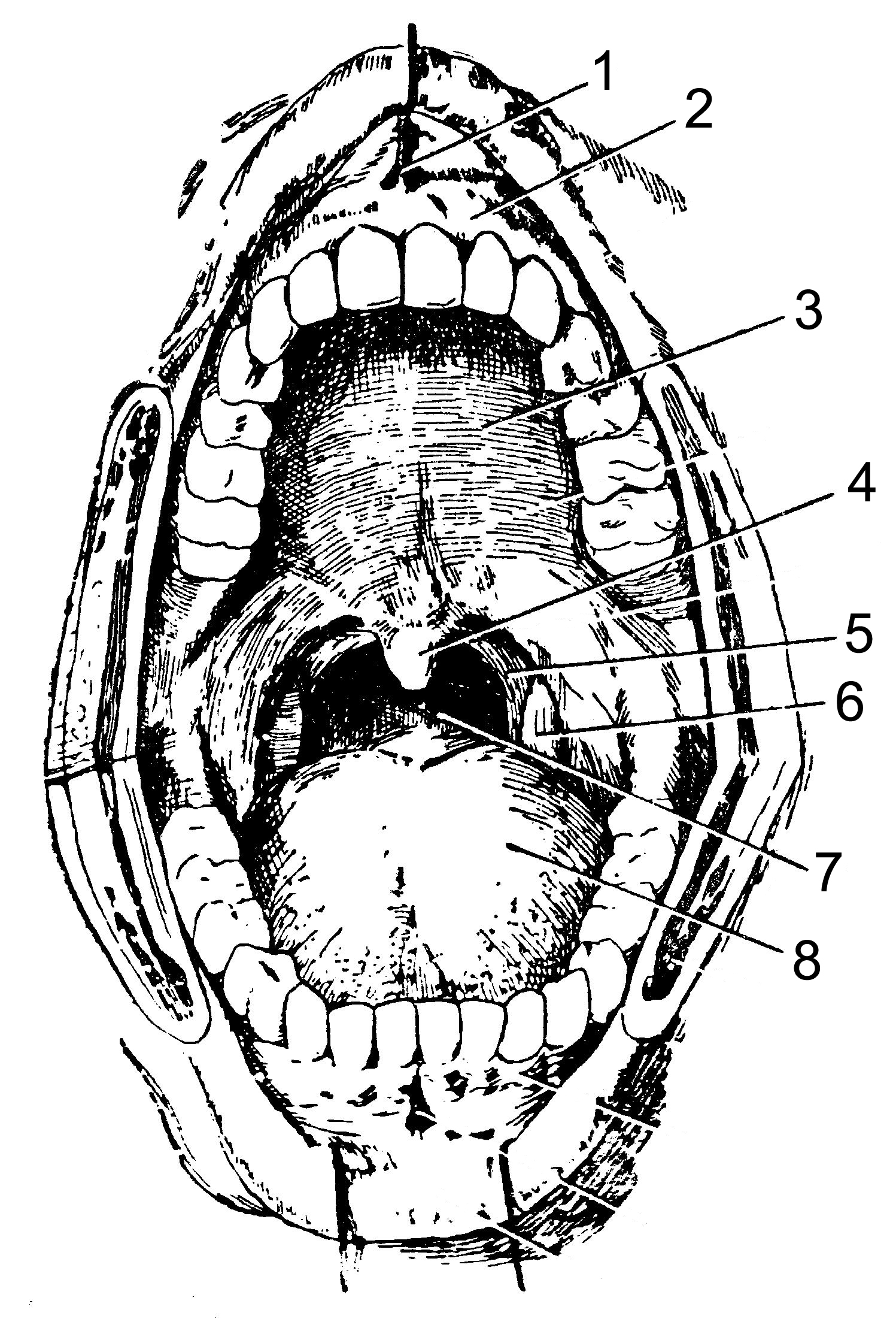
Language,lingua – lat., glossa – Greek, – a mobile muscular organ located in the oral cavity and facilitating the process of mixing food, swallowing, sucking, speech production, contains taste buds.
Structure : in the language there are:
body of tongue corpus linguae ;
the top of the tongue apex linguae ;
root of tongue radix linguae ;
back of the tongue dorsum linguae ;
edge of tongue margo linguae ;
lower surface of the tongue facies inferior linguae .
The body is separated from the root by a boundary groove, sulcus terminalis , consisting of two parts converging at an obtuse angle, at the apex of which there is a blind opening of the tongue, foramen caecum linguae .
From the lower surface of the tongue to the gums in the sagittal direction there is a fold of the mucous membrane, which is called the frenulum of the tongue, frenulum linguae . On either side of it there are paired sublingual folds, plicae sublinguales , and on them the sublingual papillae, carunculae sublinguales .
On the back and edges of the tongue, the mucous membrane is rough due to the large number of tongue papillae, papillae linguales . All papillae, except filiform and conical ones, contain taste receptors:
Filiform and conical papillae, papillae filiformes et papillae conicae , are located along the entire back of the tongue and represent a conical body with racemose appendages at the tops;
fungiform papillae, papillae fungiformes , are located on the back of the tongue closer to its edges and have the shape of pineal growths. They are larger, flattened at the edges of the tongue, their number ranges from 150 to 200;
leaf-shaped papillae, papillae foliatae , concentrated in the lateral sections of the tongue and represent 5–8 folds separated by grooves. They are unequal in size and are most pronounced in the posterior parts of the tongue;
Vital papillae , papillae vallatae , the largest, but weakly protruding above the surface, are located on the border between the root and body of the tongue, in front of the boundary line. They are cylindrical elevations surrounded by a groove around which a ridge of the mucous membrane is located. Their number ranges from 7 to 11.
The mucous membrane of the root of the tongue is devoid of papillae; under the epithelium there are lymphoid nodules, called the lingual tonsil, tonsilla lingualis .
The muscles of the tongue are represented by skeletal muscles and the intrinsic muscles of the tongue.
Skeletal muscles connect the root of the tongue to the bones of the skull:
hyoglossus muscle, m. hyoglossus , - connects the tongue to the hyoid bone. Pulls the tongue back and down;
styloglossus muscle, m. styloglossus , - connects the tongue with the styloid process temporal bone, pulls the root of the tongue up and back;
genioglossus muscle, m. genioglossus . - connects the tongue to the mental spine of the lower jaw, pulls the tongue forward and down.
Own muscles tongues have points of origin and attachment points in the thickness of the tongue, located in three mutually perpendicular planes:
inferior longitudinal muscle, m. longitudinalis inferior , shortens the tongue, lowers the tip of the tongue;
superior longitudinal muscle, m. longitudinalis superior , shortens the tongue, raises the tip of the tongue;
vertical muscle of the tongue, m. verticalis linguae , makes it flat;
transverse muscle of the tongue, m. transversus linguae , reduces its width and makes it transversely convex upward.
Glands of the mouth,glandulae oris are divided into two groups: minor salivary glands and major salivary glands:
Minor salivary glands , glandulae salivariae minores :
labial glands, glandulae labiales ;
buccal glands, glandulae buccales ;
molar glands, glandulae molares ;
palatine glands, glandulae palatinae ;
lingual glands, glandulae linguales .
Major salivary glands , glandulae salivariae majores (paired):
parotid gland, glandula paroti dea . Alveolar gland that secretes serous secretion. Located under the skin, anterior and inferior to the auricle, on the lateral surface of the branch of the lower jaw. The excretory duct opens in the vestibule of the mouth on the mucous membrane of the cheek, at the level of the upper second molar.
Submandibular gland, glandula submandibularis . The alveolar tubular gland secretes a mixed secretion. It is located in the submandibular triangle, outside the mylohyoid muscle. The excretory duct opens on the sublingual papilla.
Sublingual gland, glandula sublingualis . Alveolar tubular gland that secretes mucous secretion. It is located under the mucous membrane of the mouth on the mylohyoid muscle under the tongue. The excretory duct, together with the duct of the submandibular gland, opens on the sublingual papilla.
Figure 3 Parotid salivary gland Parotid duct Buccal muscle Masseter muscle Submandibular salivary gland Duct of the submandibular salivary gland Sublingual salivary gland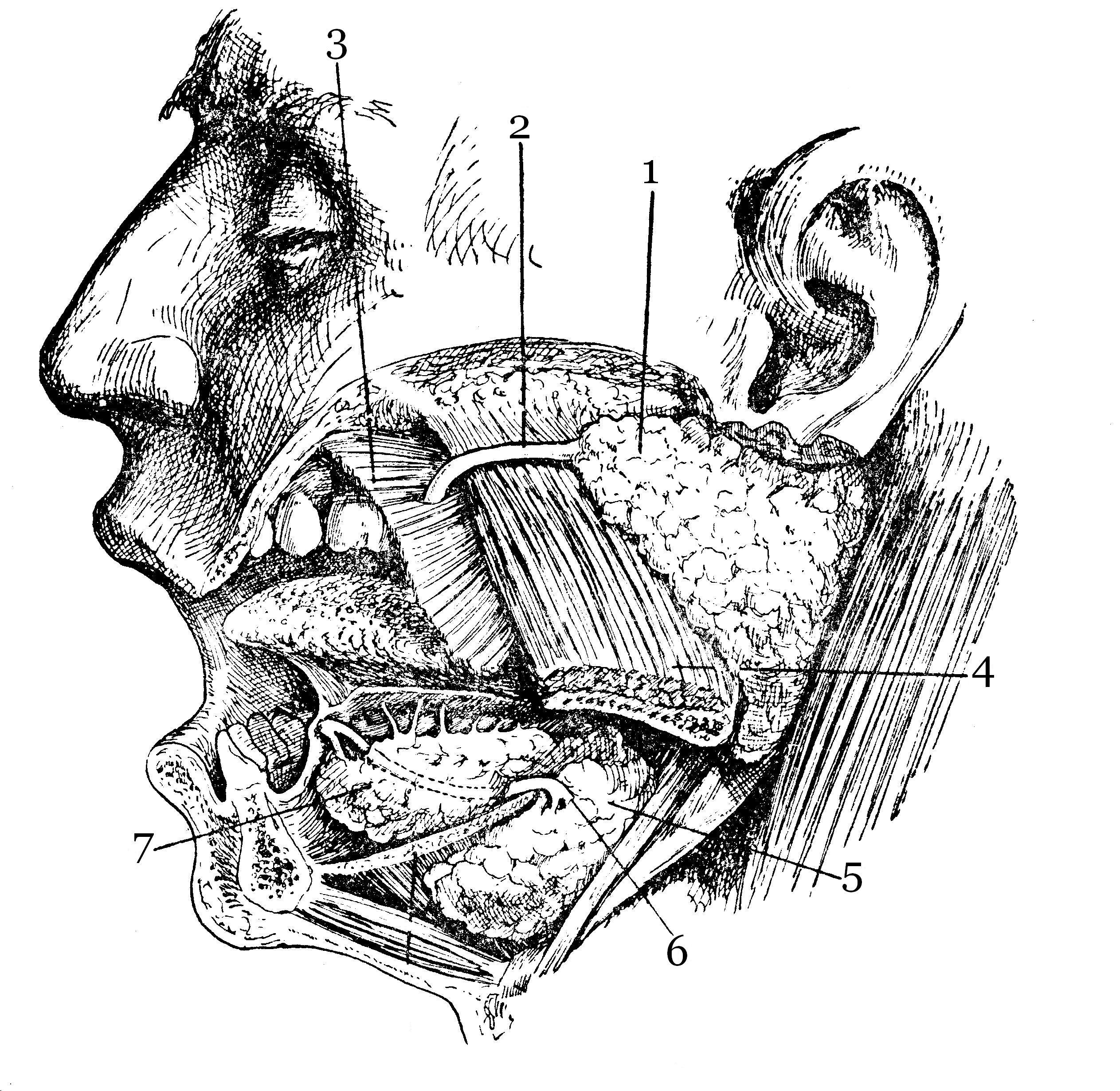
large molars, dentes molares ,
small indigenous dentes premolares ,
fangs, dentes canini ,
incisors, dentes incisivi .
Teeth,dentes , depending on their structure and functions, are distinguished:
All of them are strengthened in the dental alveoli of the upper and lower jaws. The root of the tooth and the alveolus form a continuous connection - impaction, gomphosis .
Structure:
Each tooth contains the following parts:
crown of the tooth, corona dentis , protrudes above the gum. It has lingual, vestibular, two contact and chewing surfaces;
tooth root, radix dentis . Each tooth has from one to three roots. The root ends at the apex of the tooth root, apex radi cis dentis , on which there is an opening at the apex of the tooth root, foramen apicis dentis . Through this hole into the tooth cavity containing the pulp, pulpa dentis , vessels and nerves pass through;
neck of the tooth cervix dentis , a slight narrowing of the gum;
tooth cavity, cavitas dentis . It unites the cavity of the crown, cavitas coronalis , and tooth root canal, canalis radi cis dentis .
The bulk of the tooth consists of dentin, dentinum , which is covered with enamel in the crown area, enamel um , and in the area of the neck and root - with cement, cementum . The root of the tooth is surrounded by a root membrane - periodontium, periodontium , which, with the help of tooth ligaments, attaches it to the dental alveolus.
Periods of teething:
Human teeth erupt in two periods.
In the first period, which lasts from 6 months to 2 years, the so-called baby teeth appear, dentes decidui . There are only 20 of them; on each half of the upper and lower jaws, in the direction from the median plane outward, there are 2 incisors, 1 canine and 2 large molars. Among the primary teeth there are no small molars. Dental formula: 2102.
In the second period, which lasts from 7 to 26 years, 32 permanent teeth appear, dentes permanentes . In an adult, 2 incisors, 1 canine, 2 small molars and 3 large molars erupt on each half of the upper and lower jaws. Dental formula: 2123.
Figure 4 small molars large molars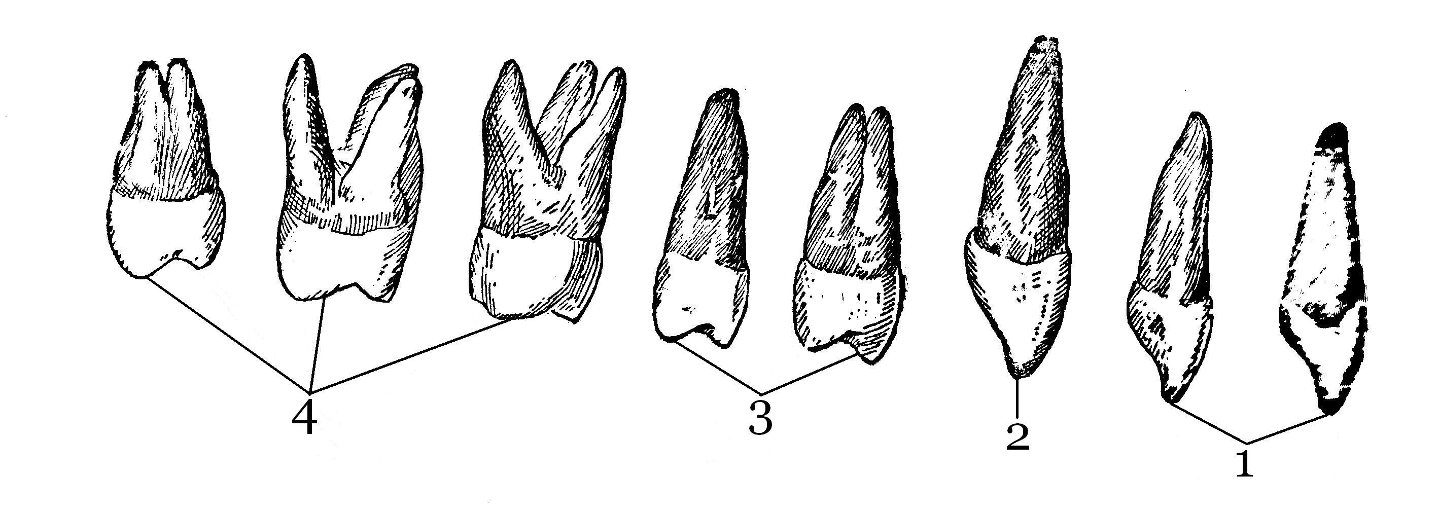
Pharynx
Pharynx,pharynx , - an unpaired organ, is a muscular tube, part of the digestive and respiratory systems.
Topography of the pharynx:
Holotopia: Located in the head and neck area.
Skeletotopia: located in front of the bodies of the cervical vertebrae from the base of the skull (pharyngeal tubercle of the occipital bone) to the level of the VI–VII cervical vertebra.
Syntopy:
at the top it is attached to the base of the skull;
behind it is the prevertebral plate of the cervical fascia, prevertebral muscles, cervical vertebrae;
from the sides – neurovascular bundles of the neck (internal jugular vein, common carotid artery, nervus vagus), large horns of the hyoid bone and plates of the thyroid cartilage;
front – nasal cavity, oral cavity and larynx.
According to the organs located in front of the pharynx, its cavity is divided into three parts: the upper - nasal, or nasopharynx, pars nasalis , middle - oral, or oropharynx, pars oralis , and the lower - laryngeal, or laryngopharynx, pars laryngea .
The nasal part is functionally the respiratory section. Unlike other sections, its walls are motionless. The anterior wall communicates with the nasal cavity through the choanae, choanae . On the lateral walls of the nasal pharynx there is the pharyngeal opening of the auditory tube, ostium pharyngeum tubae auditivae , through which the pharynx communicates with tympanic cavity. At the top and rear, this hole is limited by a pipe roller, torus tubarius . Between the pharyngeal opening of the auditory tube and the soft palate there is a paired tubal tonsil, tonsilla tubaria .
Mouth part from the front through the pharynx, fauces , communicates with the oral cavity, the posterior wall of this part corresponds to the III cervical vertebra. The intersection of the digestive and respiratory tracts occurs in the mouth.
The laryngeal part is the lower part of the pharynx, located behind the larynx and extending to the entrance to the esophagus.
Figure 5 Nasal cavity Oral cavity Solid sky Nasopharynx Oropharynx hypopharynx Laryngeal cavity Soft sky Epiglottis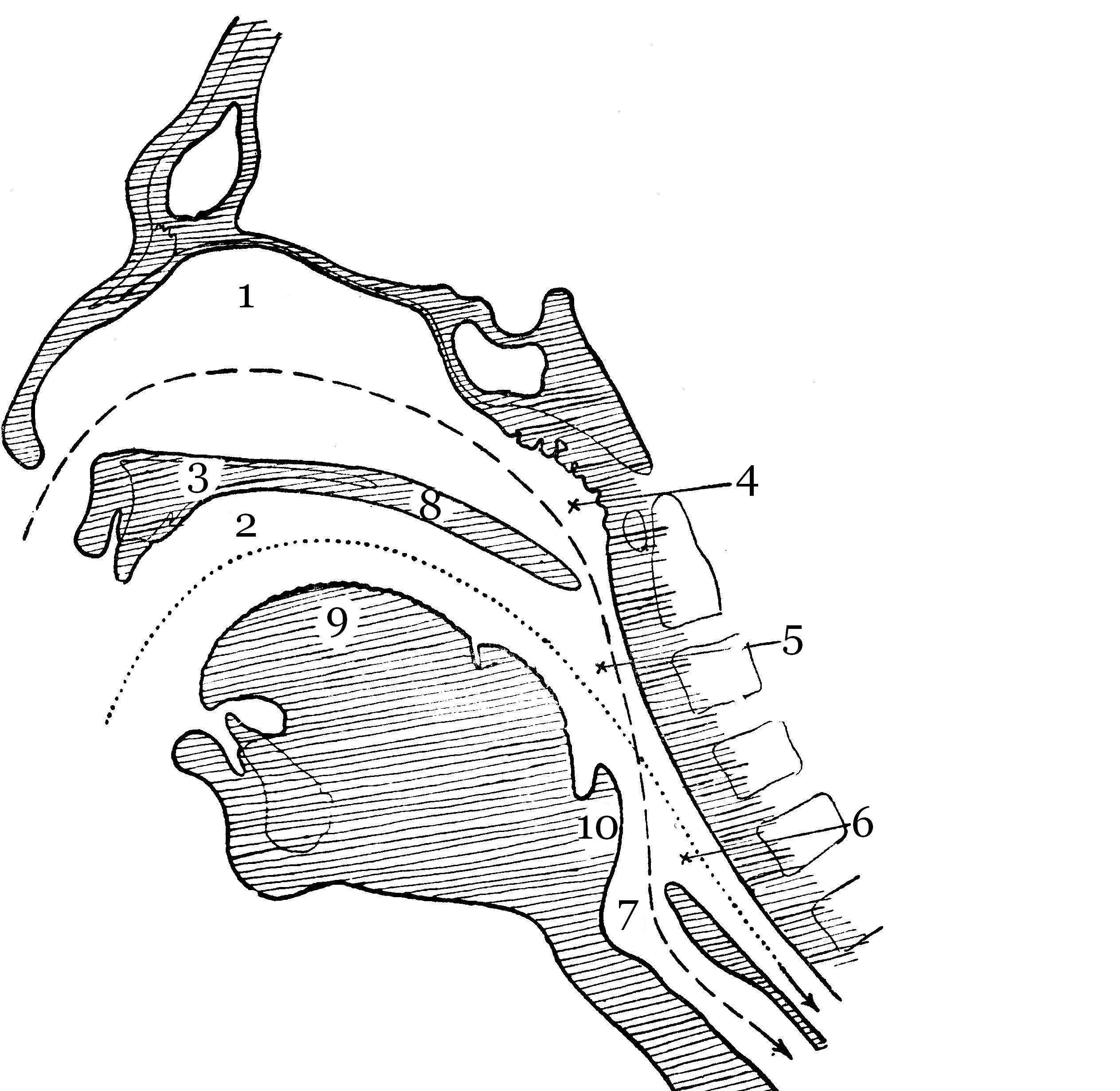
Figure 6 Upper surface of the soft palate Epiglottis Entrance to the larynx Pear-shaped pocket aryepiglottic fold Velopharyngeal arch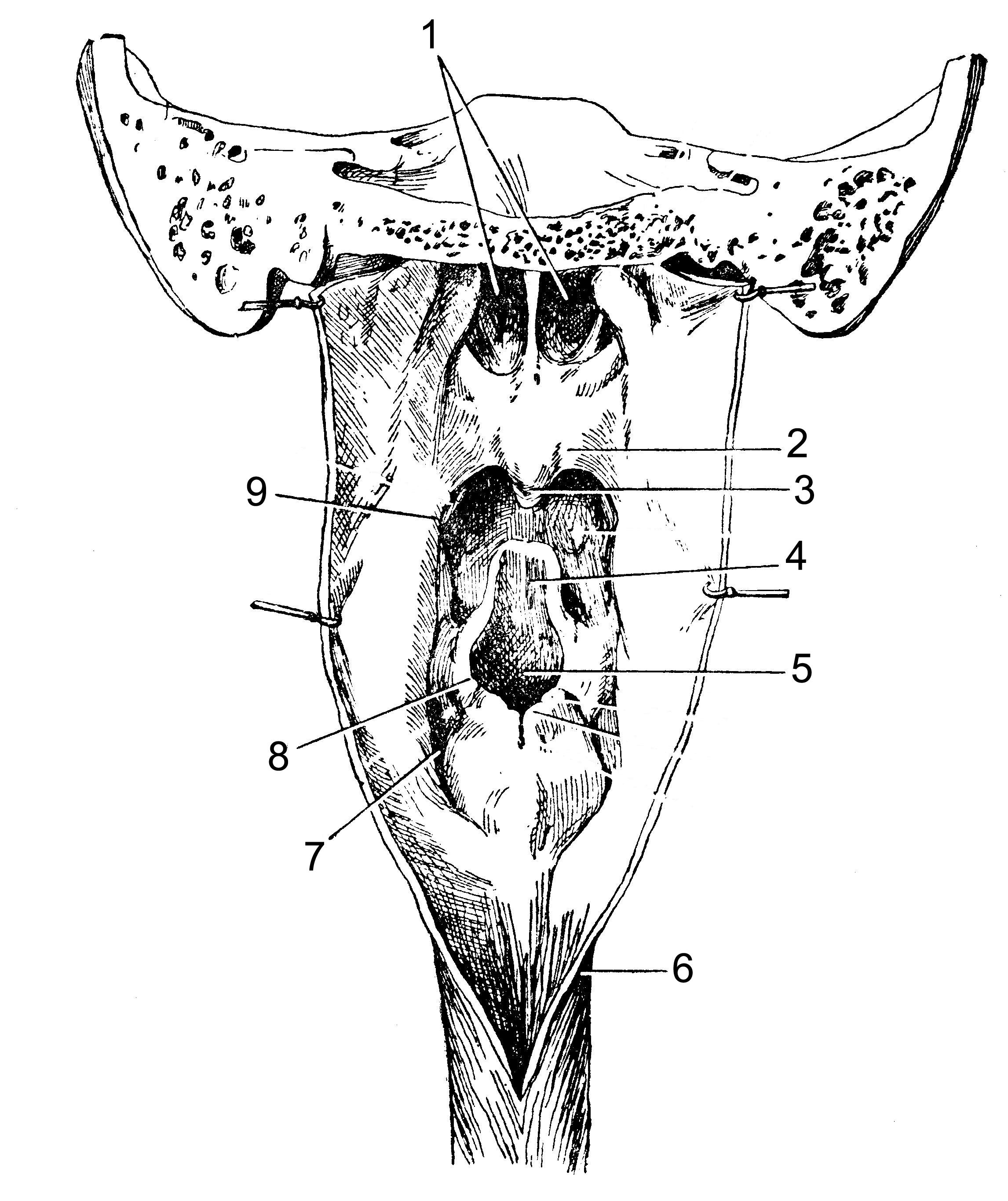
Figure 7 Superior turbinate Middle turbinate Inferior turbinate Pharyngeal opening of the auditory tube Palatolingual arch Palatopharyngeal arch Palatine tonsil Lingual tonsil Pharyngeal tonsil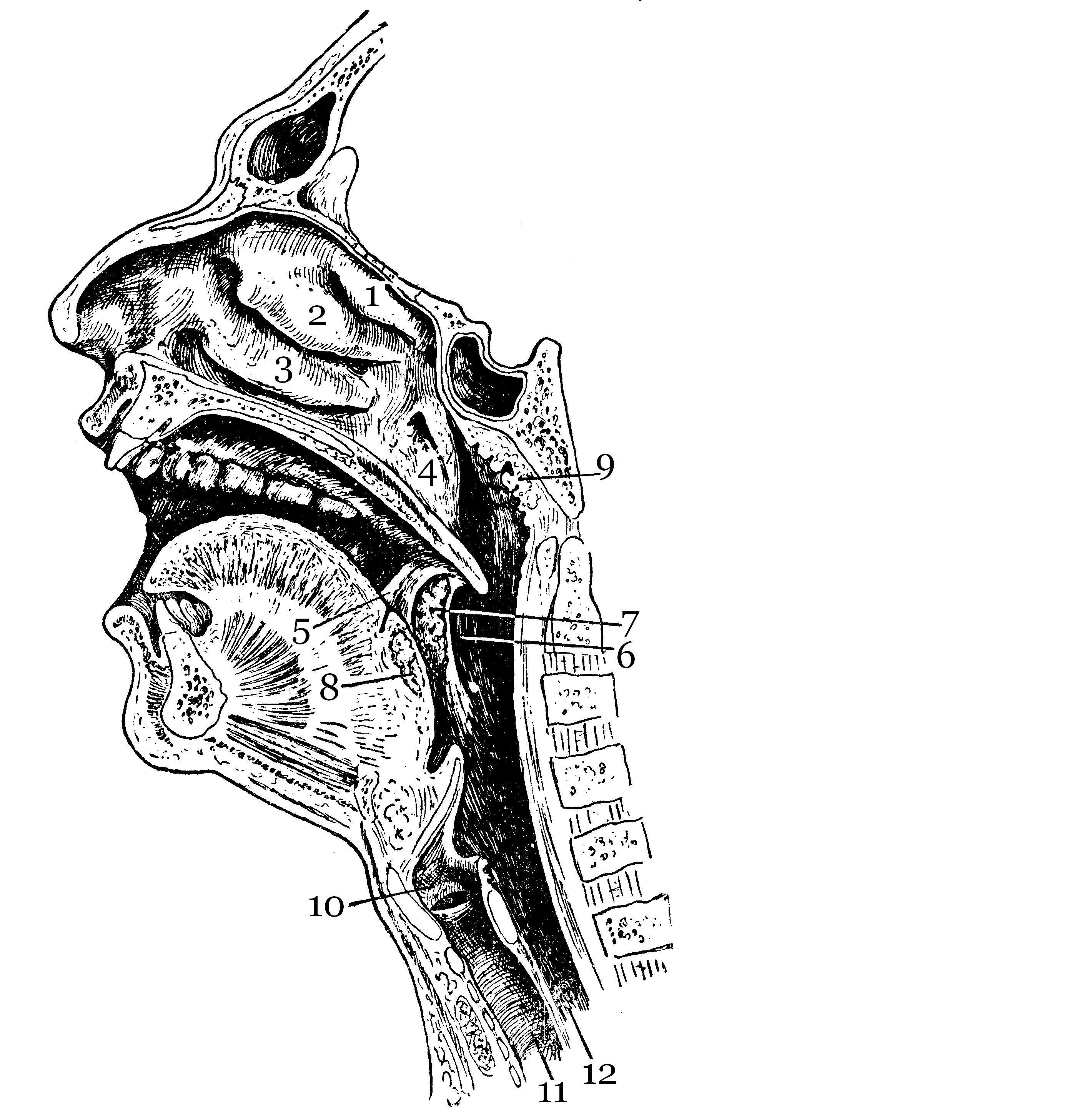
Structure of the pharynx:
The upper wall of the pharynx is called the pharyngeal vault, fornix pharyngis , and is attached to the pharyngeal tubercle of the basilar part of the occipital bone. In the area of the fornix there is an unpaired pharyngeal tonsil, tonsilla pharyng ea ( s . adenoidea ) . The internal space of the pharynx constitutes the pharyngeal cavity, cavitas pharyngis .
The wall of the pharynx consists of three membranes:
mucous membrane, tunica mucosa , The nasal part of the pharynx is covered with ciliated epithelium. In the lower sections, the epithelium is multilayered squamous. The mucous membrane lies on a connective tissue plate that replaces the submucosa. In the upper parts of the pharynx, this plate has a fibrous structure and is called the pharyngeal-basilar fascia, fascia pharingobasilaris . Starting from the oropharynx, this plate has the structure of a loose submucosal base, tela submucosa .
muscle membrane, tunica muscularis , consists of striated voluntary muscles located longitudinally (dilators) and circularly (constrictors).
The circular layer is much more pronounced and is divided into three compressors:
superior pharyngeal constrictor, m . constrictor pharyngis superior ; the upper bundles of this constrictor do not cover the wall of the pharynx in the uppermost section and, accordingly, here the wall is formed by the mucous membrane and pharyngeal-basilar fascia, covered externally with adventitia;
middle pharyngeal constrictor m. constrictor pharyngis medius ;
inferior pharyngeal constrictor, m. constrictor pharyngis inferior .
The longitudinal muscle fibers of the pharynx are part of two muscles:
stylopharyngeal muscle, m. stylopharyngeus , which raises the pharynx and narrows its lumen.
velopharyngeal muscle, m . palatopharyngeus .
Figure 8 Occipital bone Suture of the pharynx Pharyngobasilar fascia Stylopharyngeal muscle Styloglossus muscle Middle pharyngeal constrictor Hyoid bone Inferior pharyngeal constrictor Superior pharyngeal constrictor
Connective tissue membrane (adventitia), tunica adventitia is a continuation of the fascia covering the buccal muscle and passes into the connective tissue membrane of the esophagus.
Oral cavity
Oral cavity(cavum oris) (Fig. 151, 156, 194) is the beginning of the digestive apparatus. In front it is limited by the lips, above by the hard and soft palate, below by the muscles that form the floor of the mouth and tongue, and on the sides by the cheeks. The oral cavity opens with a transverse oral fissure (rima oris), limited by the lips (labia). The latter are muscle folds, the outer surface of which is covered with skin, and the inner surface is lined with mucous membrane. Through the pharynx (fauces), more precisely, the isthmus of the pharynx (isthmus faucium), the oral cavity communicates with the pharynx. The oral cavity is divided into two parts by the alveolar processes of the jaws and teeth. The anterior outer part is called the vestibule of the mouth (vestibulum oris) (Fig. 156) and is an arched gap between the cheeks and gums with teeth. The posterior internal cavity, located medially from the alveolar processes, is called the oral cavity proper (cavum oris proprium). It is limited in front and on the sides by the teeth, below by the tongue and floor of the mouth, and above by the palate. The oral cavity is lined by the oral mucosa (tunica mucosa oris), covered with stratified squamous non-keratinizing epithelium. It contains a large number of glands. The area of the mucous membrane attached around the neck of the teeth on the periosteum of the alveolar processes of the jaws is called the gum (gingiva).
Cheeks(buccae) are covered on the outside with skin, and on the inside with the mucous membrane of the mouth, which contains the ducts of the buccal glands and are formed by the buccal muscle (m. buccinator). Subcutaneous tissue is especially developed in the central part of the cheek. Between the chewing and buccal muscles is the fatty body of the cheek (corpus adiposum buccae).
Upper wall of the mouth(palate) is divided into two parts. The anterior part - the hard palate (palatium durum) - is formed by the palatine processes of the maxillary bones and horizontal plates of the palatine bones, covered with mucous membrane, along the midline of which there is a narrow white stripe, called the “suture of the palate” (raphe palati). Several transverse palatal folds (plicae palatinae transversae) extend from the suture.
Posteriorly, the hard palate passes into the soft palate (palatium molle), formed mainly by muscles and aponeurosis of tendon bundles. In the posterior part of the soft palate there is a small conical protrusion, called the uvula (Fig. 157, 195, 199), which is part of the so-called velum palatinum. Along the edges, the soft palate passes into the anterior arch, called the palatoglossus (arcus palatoglossus), which goes to the root of the tongue, and the posterior arch, the palatopharyngeus, which goes to the mucous membrane of the lateral wall of the pharynx. The palatine tonsils (tonsillae palatinae) lie in the depressions formed between the arches on each side (Fig. 152, 156, 199). The lower palate and arches are formed mainly by the muscles involved in the act of swallowing.
The muscle that strains the velum palatini (m. tensor veli palatini) (Fig. 157) is a flat triangle and stretches the anterior section of the soft palate and the pharyngeal section of the auditory tube. Its point of origin is on the scaphoid fossa, and its attachment point is on the aponeurosis of the soft palate.
The muscle that lifts the velum palatini (m. levator veli palatini) (Fig. 157) lifts the soft palate and narrows the pharyngeal opening of the auditory tube. It begins on the lower surface of the petrous part of the temporal bone and, intertwined with bundles of the muscle of the same name on the other side, is attached to the middle section of the aponeurosis of the palate.
The palatoglossus muscle (m. palatoglossus) narrows the pharynx, bringing the anterior arches closer to the root of the tongue. The point of origin is located on the lateral edge of the root of the tongue, and the attachment point is on the aponeurosis of the soft palate.
The velopharyngeal muscle (m. palatopharyngeus) (Fig. 157) has triangular shape, brings the velopharyngeal arches together, pulling up the lower part of the pharynx and larynx. Starts with back wall the lower part of the pharynx and from the plate of the thyroid cartilage, attaches to the aponeurosis of the soft palate.
Language(lingua) (Fig. 151, 152) is a mobile muscular organ located in the oral cavity and facilitating the processes of chewing food, swallowing, sucking and speech formation. The tongue is divided into the body of the tongue (corpus linguae) (Fig. 152), the apex of the tongue (apex linguae) (Fig. 152), the root of the tongue (radix linguae) (Fig. 152, 157, 195, 199) and the back of the tongue (dorsum linguae ) (Fig. 152). The body is separated from the root by a boundary groove (sulcus terminalis) (Fig. 152), consisting of two parts converging at an obtuse angle, at the apex of which there is a blind opening of the tongue (foramen caecum linguae) (Fig. 152).
From above, from the sides and partially from below, the tongue is covered with a mucous membrane, which fuses with its muscle fibers, contains glands, lymphoid formations and nerve endings, which are sensitive receptors. On the back and body of the tongue, the mucous membrane is rough due to the large number of papillae of the tongue (papillae linguales), which are divided into four groups.
Filiform papillae (papillae filiformes) (Fig. 152) are located throughout the body of the tongue and represent a conical body with racemose appendages at the tops.
Mushroom-shaped papillae (papillae fungiformes) (Fig. 152) are located on the back of the tongue closer to its edges and have the shape of pineal growths. They are larger, flattened at the edges of the tongue, their number ranges from 150 to 200.
Leaf-shaped papillae (papillae foliatae) (Fig. 152) are concentrated in the lateral sections of the tongue and represent 5-8 folds separated by grooves. They are unequal in size and are most pronounced in the posterior parts of the tongue.
The papillae, surrounded by a shaft (papillae vallatae) (Fig. 152), the largest, but weakly protruding above the surface, are located on the border between the root and the body of the tongue. They are cylindrical elevations surrounded by a groove around which a ridge of the mucous membrane is located. Their number ranges from 7 to 11.
The muscles of the tongue are represented by skeletal muscles and the actual muscles of the tongue. Skeletal muscles connect the root of the tongue with the bones of the skull: the hyoglossus muscle (m. hyoglossus) - with the hyoid bone and, together with the cartilaginous muscle (m. chondroglossus), pulls the tongue back and down; styloglossus muscle (m. styloglossus) - with the styloid process of the temporal bone, pulls the root of the tongue up and back; genioglossus muscle (m. genioglossus) (Fig. 156, 195) - with the mental spine of the lower jaw, pulls the tongue forward and down. The muscles of the tongue themselves have points of origin and attachment points in the thickness of the tongue, located in three mutually perpendicular directions: the lower longitudinal muscle (m. longitudinalis inferior) shortens the tongue; the superior longitudinal muscle (m. longitudinalis superior) bends the tongue, shortening it, and raises the tip of the tongue; the vertical muscle of the tongue (m. verticalis linguae) makes it flat; the transverse muscle of the tongue (m. transversus linguae) reduces its diameter and makes it transversely convex upward.
From the lower surface of the tongue to the gums in the sagittal direction there is a fold of the mucous membrane, which is called the frenulum of the tongue (frenulum linguae). On either side of it, at the bottom of the mouth on the sublingual fold, the ducts of the submandibular gland (glandula submandibularis) (Fig. 151) and the sublingual gland (glandula sublingualis) (Fig. 151) open, which secrete saliva and are therefore called salivary glands (glandulae salivales) . The submandibular gland is an alveolar-tubular protein-mucosal gland located in the lower part of the neck in the submandibular fossa, below the mylohyoid muscle. The sublingual gland is an alveolar-tubular protein-mucosal gland located under the mucous membrane of the mouth on the mylohyoid muscle under the tongue. The excretory duct of the third salivary gland, the parotid gland (glandula parotis) (Fig. 151), opens in the vestibule of the mouth on the mucous membrane of the cheek, at the level of the upper second molar. It is an alveolar protein gland located in the retromaxillary fossa, anterior and inferior to the external ear.
Teeth (dentes) (Fig. 151), depending on their structure and functions, are divided into large molars (dentes molares), small molars (dentes premolares), canines (dentes canini) and incisors (dentes incisivi). All of them are strengthened in the sockets of the alveolar processes of the lower and upper jaws. The method of attaching a tooth with a hole is called impacting.
Each tooth consists of a part protruding above the gum - the crown of the tooth (corona dentis) (Fig. 153), a part covered by the gum - the neck of the tooth (cervix dentis) (Fig. 153) and an internal part - the root of the tooth (radix dentis) (Fig. 153). Moreover, some teeth have two or more roots.
The bulk of the tooth is dentin (dentinum) (Fig. 153), which in the crown area is covered with enamel (enamelin) (Fig. 153), and in the neck and root area - with cement (cementum) (Fig. 153). The root of the tooth is surrounded by a root membrane - periodontium (periodontium), which, with the help of tooth ligaments, attaches it to the dental alveolus (Fig. 153). Inside the crown of the tooth, a tooth cavity (cavum dentis) is formed, which continues into a narrow canal of the tooth root (canalis radicis dentis) (Fig. 153), which opens with a small hole in the apex of the tooth root (foramen apicis radicis dentis) (Fig. 153). Through this hole, blood vessels and nerves pass into the cavity of the tooth, which contains the pulp, or pulp, of the tooth (pulpa dentis) (Fig. 153).
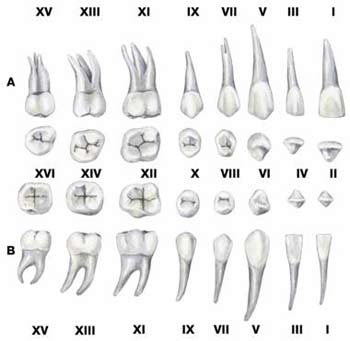
Human teeth erupt in two periods. In the first period (from 6 months to 2 years), the so-called baby teeth (dentes decidui) appear. There are only 20 of them, 10 on each jaw (Fig. 154). In the second period, which lasts from 6 to 7 years, and then from 20 to 30 (the so-called wisdom teeth), 32 permanent teeth appear (Fig. 155). In an adult, 3 large molars, 2 small molars, 1 canine and 2 incisors erupt on each half of the upper and lower jaws.
| Rice. 154. Baby teeth A - teeth upper jaw; B - teeth of the lower jaw: VI - cutting edge of the fang; VII - facial surface of the first major molar; VIII - chewing surface of the first major molar; IX - facial surface of the second major molar; X - chewing surface of the second major molar |
 |
| Rice. 155. Permanent teeth A - teeth of the upper jaw; B - teeth of the lower jaw: I - facial surface of the medial incisor; II - cutting edge of the medial incisor; III - facial surface of the lateral incisor; IV - cutting edge of the lateral incisor; V—facial surface of the canine; VI - cutting edge of the fang; VII - front surface of the first small molar; VIII - chewing surface of the first small molar; IX - front surface of the second small molar; X - chewing surface of the second small molar; XI—facial surface of the first major molar; XII - chewing surface of the first major molar; XIII - front surface of the second major molar; XIV - chewing surface of the second major molar; XV—facial surface of the third major molar; XVI - chewing surface of the third major molar |
 |
Everyone knows what the oral cavity is, but few people understand its structure. Despite its apparent simplicity, the human mouth is quite complex, and if you understand what the oral cavity itself is, you can understand the causes of many diseases.
The oral cavity is the beginning of the anterior section digestive system. It serves for receiving and primary processing of food, using different organs oral cavity. As a result, a food bolus is formed, which is sent through the pharynx into the esophagus.
The digestive functions of the oral cavity can be seen from the following table:
| Oral structure | Action | Result |
| Lips and cheeks | Retains food between teeth | · Chewing food until smooth using teeth. |
| Salivary glands | Saliva production | · Moisturizing and lubricating the mucous membranes of the mouth and throat. · Moisturizing, softening and dissolving food. · Cleaning teeth and mouth. · Salivary amylase breaks down starch. |
| External muscles of the tongue | Tongue movements to the sides, in and out | · Manipulating food for chewing. · Formation of a smooth bolus of food. · Preparing food for swallowing. |
| Intrinsic muscles of the tongue | Changing the shape of the tongue | · Preparing food for swallowing. |
| Taste buds | Feeling of food in the mouth and sense of taste | · Nerve impulses from taste buds. |
| Tongue glands | Production of the tongue enzyme lipase | · Enzyme activation in the stomach. · Breakdown of triglycerides into fatty acids and diglycerides. |
| Teeth | Tearing and crushing food | · Grinding food into small particles for grinding. |
In addition to receiving and processing food, the mouth takes part in speech communication and the breathing process. Why this happens, we will talk about this in more detail later.
How is the oral cavity limited?
The oral cavity is formed from different parts which limit it on all sides. The walls of the oral cavity are the floor of the mouth, the top and the side walls that form the palate, tongue, and cheeks.
The vestibule or vestibule of the mouth is limited internally by the teeth and gums, and externally by the lips and cheeks. IN The outer shell of the lips consists of skin, which gradually passes into the mucous membrane lining the oral cavity. The anatomy of the lips consists of tissue saturated with blood vessels, covered with a layer of keratin, which is why the lips appear red. The lips are innervated by many nerves directly connected to the cerebral cortex of the brain. This explains the special sensitivity of the lips.
The lips cover the orbicularis oris muscle, which controls jaw movement. The frenulum is a fold of mucosa located in the middle of each lip that attaches the inner surface of each lip to the gums.
The sides of the mouth are limited by the cheeks. Their outer tissue consists of skin, and the inner tissue is covered with a layer of oral mucosa. The structure of the oral mucosa (abbreviated as sopr) consists of squamous epithelium. It is arranged in layers and does not contain keratin.
A cosmetic disadvantage is the small vestibule of the oral cavity. It is corrected with an operation called vestibular deepening.
Between the skin and the epithelial mucosa of the oral cavity there is connective tissue and buccal muscles. To understand how they work, you need to pay attention to how the orbicularis oris muscles contract every time you eat, preventing food from falling back out.
With further movement deeper, you can see the opening connecting the oral cavity with the pharynx, which separates the oral cavity from the throat and is called “fauces” in Latin. Thus, the structure of the oral cavity in an anatomical sense is the area limited by the gums, teeth and fauces.
While chewing, a person needs to make an effort to breathe at the same time. To do this, the upper part of the mouth is arched, which allows you to combine chewing and breathing so that they do not interfere with each other. This arc at the top is called the sky.
What is heaven
The front part of the palate serves as a partition between the mouth and nose, as well as a solid base against which the tongue pushes food into the throat. The base of the palate contains the jaw and palatine bones of the skull, which are components hard palate. If you run your tongue along the top of the mouth, you will notice that the hard palate ends at the back of the mouth, and passes into the more “fleshy” soft palate, which consists mainly of skeletal muscles. Its soft structure allows it to change shape, which occurs involuntarily when yawning, swallowing or singing.

The uvula hangs from the posterior edge of the soft palate, located at the opening leading from the oral cavity to the pharynx. During chewing, the soft palate and uvula move forward, helping to keep food and drinks from entering the nasal cavity. The uvula also plays a role in such an annoying phenomenon as night snoring.
On the sides of the tongue there are two folds formed muscle tissue. If you look directly into the mouth, in front of the uvula you can see the palatoglossal arch, which passes through the hard palate and touches the base of the tongue at the edges. Behind the uvula there is another arch that passes through the soft palate, forming the upper and lateral edges of the fauces opening that limits the mouth.
Between these two arches are the palatine tonsils, which are formed by fused lymphoid tissue; their function is to protect the throat. The lingual tonsils are located at the base of the tongue.
What is language?
The floor of the oral cavity provides for the presence of a tongue. There is a common saying that the tongue is the strongest muscle in the human body. Those who say this do not mean absolute strength, but relative strength, which is measured in relation to size. Language is a human “workhorse” that performs many necessary tasks:
- makes swallowing easier;
- provides mechanical and chemical treatment food;
- responsible for the sense of taste (taste, texture and temperature of food);
- promotes chewing;
- provides communication through sounds.
The tongue attaches to the lower jaw at the styloid process of the temporal bone of the skull and at the hyoid bone. The floor of the mouth is formed by the mylohyoid muscles of the floor of the mouth, which move the hyoid bone. Its uniqueness is that it is located at a distance from the other bones and articulates with them indirectly.
The tongue is placed along the lower part of the mouth, forming the floor of the mouth. On the outside, the tongue consists of mucous membrane. Along its entire length stretches the middle partition (medial septum), which divides it into two symmetrical parts, each of which consists of an equal number of external and internal skeletal muscles.
The intrinsic muscles of the tongue are needed to change its shape and size. A person uses them if he wants to stick his tongue out of his mouth. They also give the tongue flexibility, which is necessary for chewing and speaking.
The extrinsic muscles originate from the outer part of the tongue and insert into the connective tissues inside the tongue. They are responsible for raising the tongue, moving it down and back, up and back, forward. All these muscles coordinate their actions with each other using nervous system and perform an important function in the process of food consumption. They place food in a position convenient for chewing, roll food into a rounded ball that is convenient for swallowing, and bring food to the edge of the mouth to make swallowing easy.
The sides and top of the tongue are densely studded with papillae different shapes, many of which are responsible for the perception of taste. The fungiform papillae have many taste buds, and the filiform papillae have tactile receptors that help the tongue move food.

Lingual glands are located in the epithelial layer of the tongue. They secrete mucus and a watery serous fluid containing the enzyme lipase. It plays an auxiliary role in the breakdown of triglycerides, but does not begin to act until it is activated when it enters the stomach.
The fold of mucous membrane on the back of the tongue is called the lingual frenulum. It attaches the tongue to the lower part of the mouth. People who suffer from a congenital condition known non-medically as tongue tie have a tongue frenulum that is too short or irregularly shaped. This disease severely impairs speech and must be corrected with cosmetic surgery.
Salivary glands
The mucous membrane of the mouth and tongue has many small salivary glands. They constantly secrete mucus either directly into the oral cavity or indirectly through the passages. The process of salivation does not stop even when a person is sleeping.
Saliva is 95.5% water. The rest is a chemical mixture of ions, glycoproteins, enzymes, growth factors and waste products. The most important component of saliva in terms of food processing is salivary amylase, which initiates the process of carbohydrate breakdown that occurs in the oral cavity. But the food does not stay in the mouth long enough for the carbohydrates to begin to be broken down. Therefore, salivary amylase continues to act until the gastric acids begin their work.
Saliva helps moisten food and makes it easier for food to move, bolus, and swallow. It contains immunoglobulin A, which prevents the penetration of microbes into the epithelium, as well as lysosomes, which give saliva antibacterial properties. Saliva contains epidermal growth factor, which has a healing effect on small wounds in the mucous membrane.
On average, each person’s body produces from 1 to 1.5 liters of saliva per day. There is usually not much of it in the mouth: no more than is needed to moisturize the mouth and teeth. The production of saliva increases during meals to moisten it and begin the chemical breakdown of carbohydrates, which occurs in the oral cavity. Small amounts of saliva are also produced by the labial glands. In addition, the glands synthesize saliva in the mucous membrane of the palate, cheeks and tongue. This ensures sufficient hydration and an adequate amount of saliva.
Major salivary glands
The glands of the oral cavity are not only small salivary glands, but also three pairs of large salivary glands, which are not part of the sorpa. They secrete saliva through salivary passages that open into the oral cavity:
- The submandibular glands are located in the lower part of the oral cavity. They secrete saliva through the submandibular salivary passages.
- The sublingual glands are located under the tongue. They use the sublingual passages to secrete saliva into the oral cavity.
- Parotid glands placed between the skin and the masticatory muscle, near the ears. They secrete saliva through the parotid canals, which exit into the oral cavity near the second upper molar.
Each of the three pairs of major salivary glands synthesizes mucus, which has special composition, inherent only to this gland. For example, the parotid glands secrete mucus that is watery in structure and contains salivary amylase. The submandibular glands have cells similar to those of the parotid glands, as well as cells that produce mucus. Therefore, their saliva, like parotid saliva, contains amylase, but not in liquid, but in thick form, diluted with mucus. The sublingual salivary glands produce the most thick saliva with the least amount of salivary amylase.
Infections of the nasal cavity and nasopharynx can spread to the salivary glands. The parotid glands are a favorite site of penetration viral infection, which causes mumps. This disease is characterized by enlarged and inflamed parotid glands, and has a characteristic appearance tumors between the ear and jaw. Symptoms of this disease include elevated temperature, a sore throat that may get worse when swallowing acidic substances such as orange juice.
How does saliva secrete?
The amount of saliva produced is regulated by the autonomic nervous system. In the absence of food, parasympathetic stimulation keeps the glands from producing saliva and maintains it at a level sufficient for comfortable speech, swallowing, sleep and others. natural processes. Salivation can be stimulated by the sight, smell and taste of food, as well as thoughts about food.
The opposite of this condition is dry mouth. This happens during times of stress, fear, anxiety. In this case, sympathetic stimulation prevails over parasympathetic stimulation and reduces saliva production. When the body is dehydrated, saliva production also decreases, causing a feeling of thirst and activity in searching for a source to satisfy it.
![]()
During eating, saliva secretion occurs as follows. Food contains substances that excite receptors on the tongue, which send nerve impulses to the superior and inferior nuclei of salivary cells in the brain stem. These two nuclei then send a signal through the parasympathetic nervous system along the fibers of the glossopharyngeal and facial nerves, which stimulate the secretion of saliva.
After swallowing food, the secretion of saliva continues for some time to clear the mouth of food debris and neutralize the irritating effect of food residues on the mucous membrane (for example, hot sauce). This saliva is mostly swallowed and reabsorbed by the body, so no fluid loss occurs.
What are teeth?
Teeth have a bony structure and are needed for tearing, grinding and grinding food. Every person has two sets of teeth - the upper arch teeth and the lower arch teeth. The first twenty baby teeth begin to emerge at six months. Between the ages of 6 and 12 years, baby teeth are replaced by 32 permanent teeth.
Each of these teeth has its own purpose:
- Eight incisors are four upper and four lower teeth. These are sharp front teeth whose job is to bite into food.
- Four canines are located on the sides of the incisors. They have a pointed tip for tearing food. These teeth are good for piercing tough meaty foods.
- To the side of the canines are eight premolars, which have a flatter surface with two prominent fang-like areas. Their function is to grind food.
- At the edge of the dental arches are 12 molars (molars), which have several fang-like projections for crushing food ready for swallowing. One of them is the “wisdom tooth”.
The teeth are fixed in the alveolar processes of the upper and lower jaw. The gum consists of soft tissues that cover and level the surface of the alveolar processes and surround the neck of each tooth. The teeth are held firmly in the sockets of the alveolar ridges by connective tissue called periodontal ligaments.
The two main parts of a tooth are the crown (the part of the tooth that protrudes above the gums) and the root, which is located deep in the upper and lower jaw. Inside they have cavities filled with pulp - soft connective tissue that contains nerves and blood vessels. The area of the pulp that is located at the root of the tooth is the root canal. The pulp cavity is surrounded by dentin, which has a bone structure. At the root of each tooth, dentin is covered with an even harder tissue - cement. In the crown of each tooth, dentin is covered with enamel, a hard shell. Enamel is the most hard fabric throughout the human body.
Although enamel protects the underlying dentin and pulp, it is susceptible to mechanical and chemical erosion, which is known as dental caries. This dental disease develops when colonies of bacteria feeding on sugars from food debris in the mouth produce acids that cause inflammation of the soft tissues of the tooth and destruction of calcium crystals in the tooth enamel.
It is worth noting that the microflora of the oral cavity is studied by oral microbiology. The microflora of the oral cavity includes more than 700 types of microorganisms. This amount is explained by the fact that the oral cavity has all the conditions for the life of microorganisms - heat, moisture and nutrients. The microflora of the oral cavity is in a state of equilibrium, when out of which microorganisms can begin to multiply intensively and cause diseases of both the oral cavity and other organs.
Throat and mouth
The throat is designed for processing food and breathing. Food enters the throat from the mouth, and air from the nasal cavity. When food enters the throat, Airways are blocked by involuntary muscle contraction.
The throat has the shape of a short tube consisting of skeletal muscles, covered inside with a mucous membrane. It extends from the back of the mouth and nasal cavity into the opening of the esophagus and larynx. The throat has three sections. The upper part of the throat (nasopharynx) is involved only in the process of breathing and the production of speech sounds. The other two sections, middle and lower (oropharynx and larynopharynx), are used for both respiration and digestion.
The lower border of the laryngopharynx connects it to the esophagus, while the anterior part of the lower throat connects to the larynx, which admits air into the trachea and respiratory tract.
The histological structure of the oropharynx is close to the structure of the oral cavity. The mucous membrane of the oropharynx consists of layers of squamous epithelium, penetrated by glands that produce mucus. During swallowing, the muscles that lift the pharynx (the digestive tube that connects the mouth and nose, as well as the esophagus and larynx) contract. At the same time, the pharynx rises and expands to accept the bolus of food. After food enters, these muscles relax, causing the muscles that compress the pharynx to force the bolus of food down the esophagus and begin peristalsis.
During swallowing, the soft palate and uvula reflexively rise to close the nasopharynx. At the same time, the larynx stretches upward, and the epiglottis, consisting of cartilaginous tissue, bends down, covering the glottis (the opening of the larynx). This process effectively closes the path for food to enter the respiratory tract, trachea and bronchi. If food or liquid goes down the wrong throat, it first enters the trachea. As a result, a cough occurs reflexively, and under the influence of convulsive movements, food is pushed out of the trachea back into the throat.
The oral cavity (cavum oris) (Fig. 210) not only serves as a place for grinding food, but is a sensitive zone where the quality of nutrients is assessed due to the receptors of general and taste sensitivity. Positive impulses that arise in the oral cavity when eating food cause an adequate response of the entire digestive system, which is expressed in the formation of digestive juices and a motor reaction corresponding to the composition of the food. Proper digestion ensures high absorption of nutrients.
The oral cavity is divided into two sections: the vestibule of the mouth (vestibulum oris) and the oral cavity itself (cavum oris proprium).
Vestibule of the oral cavity
The vestibule of the oral cavity is a narrow gap located in front between the lips and cheeks, in the back - between the upper and lower dental arches with the corresponding alveolar processes of the upper and lower jaws. Reported from external environment due to the oral fissure, and with the oral cavity itself - through the interdental spaces and the space behind the wisdom tooth. Through the last gap, a feeding probe or instrument can be inserted with the jaws closed.
In the cheek, at the level of the second upper molar, the mouth of the duct of the parotid salivary gland opens. Numerous ducts of small salivary glands also open in various parts of the mucous membrane of the vestibule.
The lips (labia oris) form the oral cavity. The upper lip reaches the septum and wings of the nose, and is limited on the side by the nasolabial groove (sulcus nasolabials). The border of the lower lip is the chin-labial groove (sulcus mentolabialis). The depth of these grooves increases with age. At the corners of the mouth, the lips are connected by adhesions. The red border and lips from the vestibule are covered with stratified non-keratinizing squamous epithelium. At the junction of the mucous membrane and the gums there are frenulums of the upper and lower lips (frenulum labii superioris et inferioris). Numerous mucoprotein glands lie in the proper layer of the mucous membrane, and the orbicularis oris muscle and the muscles that expand the oral fissure are located more superficially. Adjacent to the muscle subcutaneous tissue with sweat and sebaceous glands. The skin contains hair follicles with smooth muscles.
The cheeks (buccae), like the lips, are covered from the inside with multilayered non-keratinizing squamous epithelium. In the own layer of the mucous membrane there are small salivary and mucous glands. The muscular basis of the cheek is the buccal muscle, externally covered with a thick layer of fatty tissue (corpus adiposum buccae). The skin of the cheeks is thin and delicate, has many hair follicles, sebaceous and sweat glands.
The gums (gingivae) are a continuation of the mucous membrane of the lips and cheeks and tightly surround the necks of the teeth. Epithelial layer here it is thicker, located on a dense connective tissue basement membrane.
The oral cavity itself
The oral cavity itself is limited on the right, left and front by the upper and lower dental arches, alveolar processes, above by the hard and soft palate, below by the diaphragm of the mouth, and behind it communicates through the pharynx with the pharynx. The oral cavity contains the tongue and the sublingual salivary gland.
The oral cavity in newborns and children in the first year of life is very small: during this period of life, the jaw apparatus is underdeveloped, and the tongue fills the entire oral cavity. The cheeks are convex due to the developed fat pad of the cheeks. On the upper lip in the midline there is a tubercle connected to the gum by a short frenulum. The frenulum ends in the incisive papilla of the upper jaw. On the lower lip against the tubercle upper lip there is a depression that disappears with age. The mucous membrane also has some structural features. It is thickened, motionless in the area of the alveolar edge of the jaws and hard palate, and on the cheeks and lips it is mobile, thin and abundantly supplied with blood. There are two grooves on the alveolar edge of the upper and lower jaws: the medial one corresponds to the rudiments of milk teeth, the lateral one corresponds to permanent ones. On the wide and flattened hard palate, the mucous membrane forms transverse palatal folds.
In a newborn, the path of saliva from the mouth to the pharynx has its own characteristics. Due to the narrow vestibule of the oral cavity and the lack of a receptacle for saliva, it enters the perilingual and then the laryngopharyngeal cavity. With the appearance of molars, the parotid salivary cavity of the vestibule also appears, which communicates behind the molars with the laryngeal-pharyngeal cavity (Fig. 211).
Teeth
Teeth (dentes) are organs for grasping, biting and chewing food, participate in the articulation of speech and represent an organ of general sensitivity. They have a complex structure, origin and development. During a person’s life, as a rule, teeth grow twice: first, 20 milk teeth (dentes decidui), and then 32 permanent teeth (dentes permanentes).
Each tooth (dens) has: a crown (corona dentis), protruding into the oral cavity, and four surfaces: 1) lingual; 2) labial; 3) surface of contact with adjacent teeth; 4) chewing surface, neck (collum dentis), covered by the gum. The root (radix dentis) is held in the dental cell of the jaw by connective tissue - periodontium (periodontium).
The tooth consists of a modified bone tissue- dentin (dentinum), on the crown covered with enamel (enamelum). The dentin in the area of the neck and root of the tooth is covered with cementum.
In the center of the tooth thickness there is a crown cavity (cavum coronale) and a tooth root canal (canalis radicis dentis), which opens with an opening (for. apicis dentis) at the apex of the tooth. All this is combined into a tooth cavity (cavum dentis), filled with dental pulp (pulpa dentis), consisting of connective tissue, blood vessels and nerves (Fig. 212, 213).
Dentin is a calcified tissue. The composition of dentin includes organic substances (28%) in the form of precollagen and collagen fibers, which are impregnated with an intermediate inorganic substance (72%). In the outer layer of dentin, the fibers have a radial orientation, in the inner layer, at the border with the pulp, they have a tangential orientation. Dentin is penetrated by tubules; they contain processes of odontoblasts, the bodies of which are located in the dental pulp. The tubules at their beginning are 5 µm wide; towards the enamel border they narrow to 1 µm. With age, narrowing of the tubules is observed due to the deposition of new layers of the intermediate substance of dentin.
The enamel covering the crown of the tooth has White color with a yellowish tint. Contains little organic matter(about 3.5%) and many inorganic (96.5%); which gives greater hardness to the tooth. The enamel consists of prisms, derivatives of enameloblasts, located perpendicular to the dentin surface. The basis of the enamel is made up of thin (100 nm) reticular fibers. There is an assumption that these are calcified layers of enamel, not fibers. On the outside, the enamel is covered with a structureless cuticle, which wears off chewing surface with age.
Enamel and dentin, like all living tissues, are rebuilt throughout life. Dentin tubules are constantly washed by liquid that comes from the enamel and pulp of the tooth. With age, these processes weaken significantly.
Cement covers the root and neck of the tooth with a thin layer. By chemical composition it resembles a bone. Formed by collagen fibers impregnated with an intermediate substance, but devoid of blood vessels.
The dental pulp contains odontoblasts, fibroblasts and connective tissue fibers with a large amount of gelatinous intercellular substance. The cells produce the ground substance surrounding the pulp. The predominant cells of the pulp are fibroblasts; with age their number decreases. Other pulp cells, odontoblasts, are more differentiated cells than fibroblasts. Due to their high differentiation, odontoblasts regenerate poorly when damaged. Odontoblasts form the dentin of the tooth. Near the blood vessels of the pulp there are single histiocytes and lymphoid wandering cells. The pulp contains many reticular fibers that penetrate the dentin. Collagen fibers are collected in bundles. As a person ages, the pulp reveals larger number collagen fibers than in youth. At any age, the apical pulp contains more collagen than the coronal pulp. The main substance of the pulp is represented by proteins and mucopolysaccharides. The permeability of substances and the rate of calcification of dentin and enamel depend on the state of the basic substance of connective tissue. Therefore, if there is a deficiency of vitamins, especially vitamin C, hormones, proteins, inflammatory processes the permeability of blood vessels and the main substance of the connective tissue of the pulp is disrupted, which disrupts the function of fibroblasts and odontoblasts.
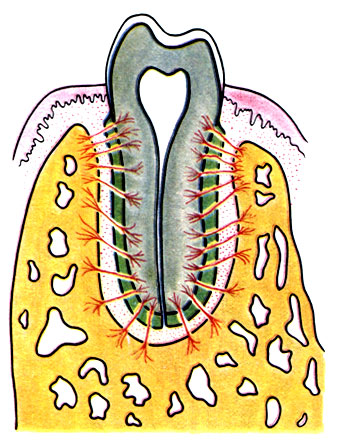
Periodontium
Dental roots are secured in the bony alveoli of the upper and lower jaws by a layer of connective tissue - periodontium, which not only holds the tooth, but also has shock-absorbing properties during loads, such as chewing.
The thickness of the periodontium ranges from 0.14 to 0.28 mm. The periodontium consists of collagen and elastic connective tissue fibers oriented perpendicularly from the walls of the alveoli to the cementum of the tooth root (Fig. 214). Loose connective tissue and its cellular elements lie between the fibers. The periodontium is well supplied with blood due to the arteries that feed the gums and is innervated. When chewing and strong clenching of the jaws, each tooth that has an antagonist is immersed in the alveoli of the jaws by 0.2 mm; in the absence of pressure, the teeth return to their original position due to the elasticity of the periodontium. With age, its elasticity and thickness decrease, which leads to a large discrepancy between the tooth root and the bone alveolus. For vitamin deficiencies (lack of vitamin C) and poisoning of the body (mercury, fluorine, salts heavy metals), during periodontal inflammation, connective tissue fibers are destroyed and teeth fall out.
Baby teeth
There are 20 primary teeth. Their size is 35% of the size of permanent teeth. They have a relatively well-developed crown and neck, short and thin roots. Milk teeth are represented by incisors, canines, large molars; each of them has certain periods of eruption and change. They are designated:
Timing of eruption of primary teeth Medial incisors.................6-8 months. Lateral...................7-8 months. Anterior molars............12-15 months. Fangs........................15-20 months. Posterior molars..............20-24 months.
Permanent teeth
There are 32 permanent teeth. There are incisors (dentes incisivi), canines (dentes canini), small molars (dentes premolares), large molars (dentes molares) (Fig. 215). They are designated:

The replacement of baby teeth with permanent ones occurs in the following sequence.
First molar.............6-8 years Medial incisors................6-9 years Lateral incisors...................7-10 years First small molar...................9 -13 years Fang...................................9-14 years Second small molar.. .............11-14 years old Second molar................10-14 years Third molar........ .....18-30 years (wisdom teeth - unstable)
Signs of teeth. Signs of teeth are used to distinguish the teeth of the same name in the right and left dental arches. When the teeth are positioned in the vestibular norm, three signs are distinguished: 1. Crown angle sign - the angle formed by the chewing and medial surfaces of the crown is sharper than the rounded angle between the chewing and lateral surfaces.
2. The sign of enamel curvature is determined on the crown from the chewing surface. The lateral part on the vestibular side is more convex.
3. The root sign is determined by the deviation of the longitudinal axis of the tooth in relation to the longitudinal axis of the crown. The longitudinal axis of the crown is projected from the middle of the cutting edge perpendicular to it, and longitudinal axis The tooth is drawn from the apex of the root to the middle of the cutting edge. In this case, the deviation of the direction of the longitudinal axis of the tooth indicates the side of the tooth.
Incisors (dentes incisivi). There are 2 upper and 2 lower medial incisors, 2 upper and 2 lower lateral incisors. The crown has the shape of a chisel with a cutting edge. In young people, three tubercles are localized on the cutting edge, which wear out with age. The labial surface of the crown is convex, lingual - it has a pronounced single tubercle at the junction of the crown and the neck. The largest crown is on the medial incisors. The single root of each tooth (rarely there are two) is round in shape and tapers conically at the apex.
The root sign is characterized by the fact that the longitudinal axis of the tooth intersects in the middle a line running parallel to the cutting edge, and not perpendicular; the result is a larger angle open to the midline and jaw. The sign of the angle is based on the fact that the medial angle is acute or straight, and the lateral angle is more than 90°. The sign of enamel curvature emphasizes the different curvature of the labial surface of the tooth; it is convex at the medial edge and flattened at the lateral.
Fangs (dentes canini). There are 2 fangs on the upper and 2 on the lower jaw. They are located on the outside of the lateral incisors. The crown is cone-shaped, the labial surface is more convex, the lingual surface is flattened, and has a tubercle. The roots of the canines are longer than those of the incisors and are compressed from the sides. The upper canines have longitudinal indistinct grooves and are better developed than the lower ones. To distinguish between right and left teeth, there are signs of root, angle and curvature. In addition, the location of the enamel border helps in determining: on the lingual surface it has an arched line, on the medial surface it rises to the crown, and on the lateral surface it descends to the root.
Primary canines are characterized by a more clearly defined sharp cone of the crown and longitudinal ridges on the labial and lingual surfaces.
Small molars (dentes premolares). In total there are 4 upper and 4 lower, located behind the fangs. They are designated as the first and second molars. The shape of the crown and root of these teeth differs from all previous ones. The chewing surface contains buccal, more pronounced, and lingual tubercles. The teeth of the upper jaw have more prominent tubercles. There is a ridge on the chewing surface of the first tooth between the tubercles; On the sides of it there are pits, deeper at the buccal tubercle. At the second tooth, on the side of the lingual tubercle, there is an incomplete groove, forming two minor elevations.
The upper teeth have a flattened root, sometimes forked at the end; The root of the lower teeth is always one, cone-shaped.
Large molars (dentes molares). There are a total of 6 teeth on the upper and 6 teeth on the lower jaw; are located behind the small molars. The third tooth is the wisdom tooth (dens serotinus).
The crown of the large molars of the upper jaw has rounded corners, which creates an irregular diamond shape. On the chewing surface, 2 buccal and 2 lingual tubercles are visible, separated by deep grooves. The exception is the second large molar of the upper jaw, where an additional tubercle (tuberculum anomale carabelli) occurs. The tubercle is well defined in apes. The presence of an underdeveloped similar tubercle in a human tooth confirms its evolutionary origin. These teeth have two buccal roots and one lingual (palatal) root. The posterior buccal root is shorter. Often the roots reach the bottom maxillary sinus.
The crown of the lower molars is shaped like a cube and has several big sizes than upper teeth. The lingual and anterior surfaces of the crown are flat, while the buccal and posterior surfaces are raised. The first large molars of the lower jaw often have 5 tubercles on the chewing surface: 3 buccal and 2 lingual, the second and third have 4 tubercles each. The lingual cusps are sharper than the buccal ones.
Each tooth has a wider root at the front; the posterior root is narrow, without a groove. The third large molar is poorly developed: its crown and root sizes are smaller, and the number of roots is not constant and ranges from 1 to 5.
Intraoral dental radiographs
(X-ray data are presented according to L. D. Lindenbraten (1971).)
The image examines the crowns and necks of teeth, roots of teeth, periodontium, compact bone plate of the alveoli, spongy bone substance of the alveolar process, and the alveolar margin.
An intraoral radiograph must meet three requirements: a separate image of the teeth being examined, no deformation of their shadow, a clear image of the structure of the edge of the lower jaw.
Intraoral radiographs provide images of a small area of the jaw. In the photographs of the upper jaw, its structure is represented by a finely looped pattern with a different arrangement of bone beams, and the nasal cavity and maxillary sinus are also visible. Images of the lower jaw reveal a large-loop bone structure with clearly visible horizontal struts, and the mandibular canal is often visible.
The photograph of the upper central incisors shows the anterior part of the nasal cavity, the shadow of the nasal septum and the anterior nasal spine (Fig. 216). An oval or rounded incisive opening appears between the roots of the central incisors or above them. The crown of the central incisors is spade-shaped, wider than that of the lateral incisors. Outside the lateral incisor, a canine can be seen, which has a triangular spear-shaped crown and a longer root than the other teeth.
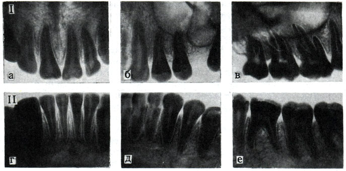
The photograph of the small molars of the upper jaw shows a continuous shadow pattern. If you carefully examine the image, you can distinguish the bottom of the maxillary sinus, outer wall nose and the edge of the zygomatic bone. Small molars have two cusps on the chewing surface. The first small molar has two roots (buccal and palatal), the second has one. The image of the large molars of the upper jaw also reveals the bottom of the maxillary sinus and cheekbone, and behind the last large molar a shadow of the maxillary tubercle is found. Large molars have wide crowns with several chewing cusps; the first two large molars have three roots each - two buccal (medial and distal) and palatal; the latter appears longer in the image due to projection distortion.
On photographs of the teeth of the lower jaw, relatively few details are noted in the area of the incisors - the shadow of the mental tubercle in the area of the small molars and the mental foramen. The lateral incisors are wider than the central ones; the fang has a spear-shaped crown and the longest root.
The small molars have two chewing cusps. The lower molars have large crowns with several chewing cusps, medial and distal roots. Below the roots, the mandibular canal appears as a clearing. Affected teeth are marked according to a generally accepted formula.
On radiographs, the periodontium appears as a dark strip 0.2-0.25 mm wide; interalveolar septa reach the necks of the teeth.
Dental bite
The teeth of the upper and lower jaws form dental arches. The upper arch is wider and covers the lower arch, located anterior and outward from it. The closure of the dental arches in relation to one another is called occlusion. There are central, anterior, right and left occlusion. The relationship of the dental arches in central occlusion is called occlusion. Physiological bites have different shape: a) orthognathia - the teeth of the upper jaw slightly overlap the teeth of the lower jaw; b) progeny - the teeth of the lower jaw overlap the teeth of the upper jaw; c) biprognathia - the teeth of the upper and lower jaws are tilted forward with partial overlap of the lower teeth by the upper ones; d) straight bite - the cutting edges of the upper and lower teeth correspond to each other.
Naturally, in practice there are many pathological variants of occlusion, depending on the incorrect formation of the dental arches and jaws.
Phylogeny of teeth
The teeth were previously scaly skin formations, which along the edges of the mouth opening developed so much that they rose above the epidermis and became capable of capturing and holding food. Due to the nature of the food, the shape of the teeth also changed. Initial and simplest form teeth - conical. Conical teeth are numerous and are strengthened by accretion to the bone or grow into it, or are located in the thickness of the oral mucosa.
Selachia have numerous conical teeth, sometimes connected into flat large plates, identical in structure to placoid scales skin. Confirmation that these plates are a derivative of numerous conical teeth is their internal structure, where numerous canals are visible. In the oral cavity, during movements of the jaw apparatus and grasping food, placoid scales differentiated into dense conical teeth. Bony fish have more than 200 teeth in the oral cavity, scattered over the entire surface of the mucous membrane; they even occur in the pharynx. Amphibians have many small teeth throughout the mouth.
Reptiles have conical teeth, attached to the inner surface and along the edges of the jaws. Some teeth sink into the alveoli with their roots. Poisonous snakes have teeth that are hollow or have grooves connected to the ducts of the poisonous glands. When bitten, the poison flows through the canal or groove of the tooth into the wound. A characteristic feature of reptiles is an unlimited number of teeth changes.
Some mammals have 44 teeth located in the jaws. Each tooth has a deep cavity in the bone. The teeth were differentiated into incisors (for grasping and cutting food), canines (for tearing food) and molars (for grinding food). In apes and humans, the number of teeth has decreased to 32. Mammals have only two changes of teeth, and small molars do not have milk teeth at all. The shape of the crown in animals is very diverse and depends on the quality of the food consumed.
Development of teeth in embryogenesis
Tooth enamel is a derivative of the epithelium, and dentin is a connective tissue layer of the skin. This confirms the comparative anatomical data according to which teeth are a derivative of placoid skin scales.
At the 7th week of the embryonic period, thickenings of the epithelium - dental plates - appear along the edges of the upper and lower jaws. At week 8 intrauterine development they grow into the underlying mesenchyme of the jaws, where protrusions are formed to form the enamel crowns of milk and permanent teeth (Fig. 217). In the enamel crown of the teeth there are cylindrical cells - adamantoblasts, which form the enamel of the teeth. In the inner part of the enamel organ there is mesenchyme, which differentiates into the dental papilla, where the outer cells turn into odontoblasts, which secrete an intermediate substance and form the fibrous formations of dentin. The pulp develops from the inner part of the dental papilla.
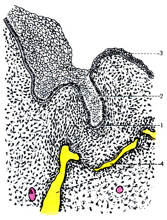
At the 19th week of the embryonic period, the dental plate is completely separated from the epithelium of the oral cavity. Odontoblasts form numerous processes that penetrate dentin, passing through special dentinal tubules. Odontoblasts form the organic and inorganic substance of the tooth. When odontoblasts are destroyed, dentin gradually darkens, becomes more fragile and is destroyed.
On the border between outer surface In the developing tooth bud and bone, an accumulation of mesenchymal cells in the form of a dental sac is observed. During teething, the sac under the crown is destroyed, and the remaining part of it fits tightly to the dentin of the root, forming the periosteum. It contains cementoblasts that form the intermediate substance and form cement fibers.
Between the root of the tooth and the bone there is a space filled with fibrous connective tissue, which turns into fibrous ligaments. These fibers are immured with cement on the side of the tooth root, and with bone substance on the side of the jaw bone. Thus, periodontal connective tissue fibers are fixed in the tooth and bone.
Changing teeth
For IV-V months. During intrauterine development, the enamel organ of permanent teeth, similarly to milk teeth, arises from the dental plate and is located on the lingual side next to the milk teeth (Fig. 218). Simultaneously with the growth of permanent teeth, the roots of baby teeth are destroyed, and their crowns fall off. A new permanent tooth appears in place of the lost tooth.
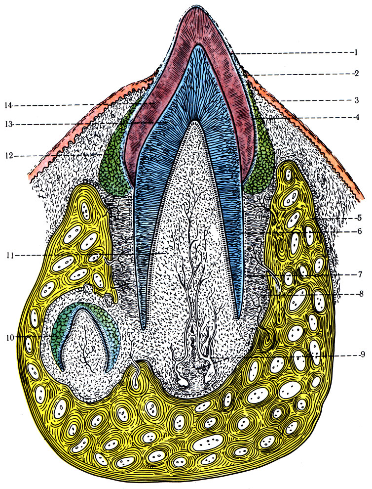
Anomalies of dental development
A common anomaly is the presence of an additional incisor or fourth molar. Quite often there is also the absence of a third large molar, which erupts with a significant delay only after 18 years. Sometimes wisdom teeth erupt after 30-40 years. If there is no wisdom tooth, the second large molar is also underdeveloped: it has three tubercles instead of four and a small crown. In some cases, there is underdevelopment of the lateral incisors, second small molars or their complete absence.
Some teeth are located not along the line of the alveolar process, but on the lingual or buccal side of the alveolar arch, which can easily be corrected using orthopedic methods during teething.
Regarding the number of roots, there are options: the roots of the tooth merge into one or, conversely, split into two or three; individual roots bend like a hook or diverge radially.
In diseases and impaired metabolism, the processes of calcification of bones and teeth may be disrupted, as well as changes in the formation of fibrous structures. These disorders often do not appear in the bones; they are clearly visible in the enamel and dentin of the tooth, where, due to improper deposition of the intermediate substance and impregnation with pigments, dark spots form, small and large cavities appear, and toothache occurs due to pressure.
Sky
The palate (palatum) forms upper wall the oral cavity and consists of the hard palate (palatum durum) and the soft palate (palatum molle).
Solid sky
The hard palate is represented by the palatine processes of the maxillary bones and horizontal plates of the palatine bones, which are connected to each other by sutures. The shape of the hard palate can be different, but in general it has the appearance of a dome-shaped plate, which is covered with a mucous membrane. Behind the incisors, on the sides of the median suture, the mucous membrane forms 2-5 transverse ridges. In the area of the incisive foramen, sometimes there is a thickening of the mucous membrane - the incisive papilla. The mucous membrane is covered with stratified non-keratinizing squamous epithelium. The submucosal layer thickens and forms a fibrous plate that fuses with the periosteum. The fusion is especially strong in the area of the sutures and at the transition to the gums, so the mucous membrane of the hard palate is motionless. In other places, between the lamina propria of the mucous membrane and the periosteum, a thin layer of adipose tissue is localized, in which small mucous palatine glands (gll. palatinae), which have a tubular-alveolar structure, are located.
Soft sky
The soft palate is attached by its anterior edge to the posterior edge of the hard palate. At the back, it ends with a velum palatine with a uvula in the middle, separating the nasopharynx from the oropharynx.
The soft palate is a muscular aponeurotic formation covered with a mucous membrane. On the side of the oral cavity, the mucosa is covered with stratified non-keratinizing epithelium, and on the side of the nasopharynx - with multi-row ciliated epithelium. Multilayered squamous epithelium is located on a well-developed basement membrane with a large number of elastic fibers, and in the thickness of the basement membrane of the mucous membrane with ciliated epithelium there are numerous mucous glands, the secretion of which moisturizes the surface of the mucosa.
In the lateral sections of the soft palate there are two arches, covered with a mucous membrane (arcus palatoglossus et palatopharyngeus), in the thickness of which the muscles of the same name are located. Between the arches there is a depression (sinus tonsillaris), where the palatine tonsil is located.
The basis of the soft palate is formed by muscles and their tendons (Fig. 219).
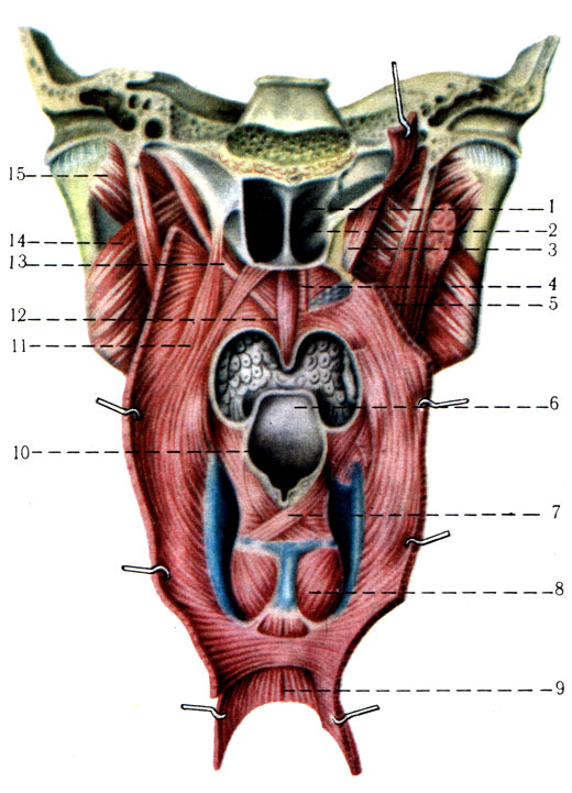
1. The muscle that strains the velum palatini (m. tensor veli palatini), paired, lives up to its name. Starts from the cartilaginous part of the auditory tube of the middle ear, from the base and medial plate pterygoid process and spina angularis sphenoid bone, then follows down and reaches the uncinate process of the medial plate, where a thin tendon is thrown over the hook, heading up and medially. Having reached the soft palate, the muscle tendon fan-shapedly diverges in the form of an aponeurosis, which connects with a similar aponeurosis on the opposite side. This tendon forms the basis of the soft palate.
Innervation: n. tensoris veli palatini.
Function. Pulls the velum and can partially lower it.
2. The muscle that lifts the velum palatini (m. levator veli palatini) is paired. This muscle is more developed than the previous one. Starts from the lower surface of the pyramid of the temporal bone between for. caroticum externum and the cartilaginous part of the auditory tube, follows down and medially, ending in the velum palatine.
Function. Raises the soft palate.
3. The palatoglossus muscle (m. palatoglossus) is paired, in the form of a thin plate, located in the fold of the mucous membrane of the same name. It starts from the aponeurosis of the soft palate, descends to the tongue and at its root connects with similar bundles of the opposite muscle.
Function. Lowers the soft palate, narrows the exit of the oral cavity into the pharynx.
4. The velopharyngeus muscle (m. palatopharyngeus) is paired, located in the thickness of the fold of the mucous membrane of the same name, located somewhat posterior to the arcus palatoglossus. It starts from the aponeurosis of the soft palate, then passes down and weaves into the back wall of the pharynx.
Function. Lowers the soft palate, reduces the entrance to the pharynx.
5. The uvula muscle (m. uvulae) is not paired, weak and small. It starts from the aponeurosis of the soft palate, and then descends to the top of the uvula and is woven into the mucous membrane.
Innervation: All four muscles receive branches from the plexus pharyngeus.
Function. Pulls up the top of the tongue.
Thus, the soft palate, consisting of mucous membrane and muscles, changes its position. When a bolus of food passes from the mouth, the soft palate rises and tightly isolates the oropharynx from the nasopharynx. The soft palate takes part in the act of breathing and speech.
Isthmus of pharynx
The isthmus of the pharynx (isthmus faucium) (Fig. 210) is an opening that connects the oral cavity with the pharynx. On the sides, the pharynx is limited by paired arches (arcus palatoglossus, arcus palatopharyngeus), in the thickness of which there are muscles of the same name. The arches limit the fossa for the tonsil (fossa tonsillaris). Below, the isthmus of the pharynx is limited by the root of the tongue, above by the lower edge of the soft palate.
Language
The tongue (lingua s. glossa) consists of striated muscles covered with mucous membrane. The importance of language is enormous. In the mucous membrane of the tongue there are many sensitive (taste) nerve receptors, general sensitivity receptors that evaluate physical properties food (hot, cold, hard, dry, wet), autonomic nerve endings (sympathetic and parasympathetic) innervating the smooth muscles of the walls of the vessels of the tongue, and numerous mucous glands. Irritation when eating taste buds and general sensitivity receptors causes swallowing, muscle contraction gastrointestinal tract and juice separation. The tongue, as a muscular organ, is actively involved in performing complex movements when grasping, mixing food and evacuating it into the pharynx. Together with the teeth and lips, it participates in the formation of articulate sounds (speech).
The tongue is conventionally divided into three parts: the tip (apex) - the free part, the body (corpus), the root (radix) - between the blind foramen and the hyoid bone. Its upper surface - the back (dorsum) - is free throughout its entire length (Fig. 220).
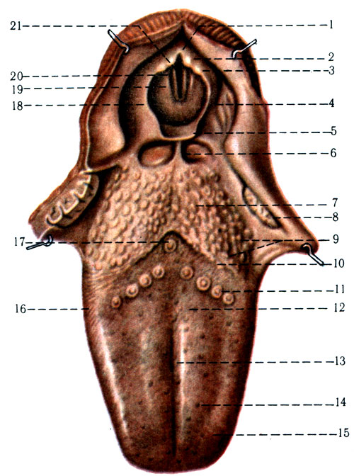
The mucous membrane on the lower surface of the tongue is covered with stratified non-keratinizing epithelium. On the back of the tongue, the mucous membrane forms outgrowths - papillae, which have different sizes and shapes.
Filiform (papillae filiformes) and cone-shaped (papillae conicae) (Fig. 221) papillae make up the majority of all papillae of the tongue. They have the shape of cut threads and are conically pointed. The epithelial lining at their apices sometimes becomes keratinized and can be torn off. In humans, especially with certain diseases, the process of keratinization is accelerated, and rejection is slowed down. In these cases, the tongue is covered with a grayish coating of epithelial plates. The connective tissue at the base of the papillae contains nerve receptors for general sensitivity.
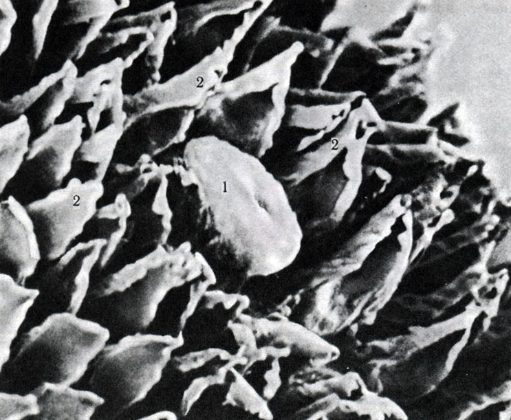
Mushroom-shaped papillae (papillae fungiformes) have the shape of a mushroom, scattered evenly along the back of the tongue. Their apex is expanded, covered with stratified non-keratinizing epithelium and rises somewhat above the other papillae. They have a light red tint and are clearly visible against the gray background of the filiform and conical papillae. At the base and expanded part of each fungiform papilla there are taste buds, where taste nerve endings lie.
The grooved papillae (papillae vallatae), numbering 7-12, form an angle, at the apex of which there is a blind opening (foramen cecum). The circumvallate papillae are located at the border of the body and the root of the tongue. Around each papilla there is a deep cleft lined with stratified epithelium, which contains taste buds and the mouths of protein glands. Nutrients dissolved in water flow into the gap of the grooved papillae and cause irritation of the taste buds until the gap is washed with the secretion of the protein glands.
Leaf-shaped papillae (papillae foliatae) are better visible on the lateral surface of the tongue in childhood in the form of 4-9 parallel elevations. They also contain taste buds.
In older people, atrophy of the leaf-shaped papillae and mucous glands of the lateral surface of the tongue occurs. In the submucosal layer of the tongue, fatty lobules appear instead.
The glands of the tongue, according to the nature of the secretion they secrete, are divided into three groups: protein, mixed and mucous. Protein glands are simple tubular glands with narrow ducts opening into the slit of the circumvallate papillae. Mixed glands are located in the root and along the edges of the tongue and have an alveolar-tubular structure. The secretory sections of the mixed glands lie in the muscles of the tongue. The ducts open under the tongue in a fold of the mucous membrane. Mucous glands are located in the mucous membrane of the back of the tongue.
Muscles of the tongue. The intrinsic muscles of the tongue are formed by striated muscles located in three mutually perpendicular planes in the thickness of the tongue. These muscles are separated by the septum linguae. The tongue has muscles connected to the lower jaw (hyoid bone).
Own muscles of the tongue. The upper and lower longitudinal muscles (mm. longitudinales superior et inferior) begin from the root of the tongue, the hyoid bone and lie at different depths, reaching the tip of the tongue.
Function. When both muscles contract, the tongue shortens; when only the upper longitudinal muscles contract, the tip of the tongue turns towards the back, and the lower ones - towards the frenulum of the tongue.
The transverse muscle (m. transversus) is oriented in the frontal plane. In the area of the tongue root it is strengthened due to m. palatoglossus.
Function. The tongue becomes narrower and longer, and also curls into a groove.
The vertical muscle (m. verticalis) starts from the lower surface of the tongue and reaches the back.
Function. Contraction of the muscle causes the tongue to flatten.
All muscles are innervated by pair XII cranial nerves. The intrinsic muscles of the tongue are located in three mutually perpendicular positions, which ensures movement of the tongue in all directions.
Muscles of the tongue starting on the skeleton: m. genioglossus, m. hyoglossus, m. styloglossus (Fig. 222); their attachment and function are described in the section on the muscles of the neck.
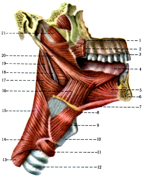
A newborn's tongue is short, wide and thick, disproportionately large in relation to the oral cavity. With the jaws closed, its edges and tip are located between the alveolar edges, touching the mucous membrane of the cheeks and lips. In a newborn and children of the first years of life, under the tongue there is a more pronounced, fringed fold of the mucous membrane than in adults. At the border of the body and the root of the tongue, a deep groove runs in the direction of the foramen cecum. The mucous membrane of the back of the tongue contains a larger number of taste bud rudiments, but significantly fewer mucous glands. In the elderly, the papillae of the tongue significantly atrophy, the mucous membrane becomes thinner.
Comparative anatomy of the tongue
In the oral cavity of fish, the tongue is represented by a fold of mucous membrane. There are no muscles in it and its movements are carried out together with the ventral part of the gill apparatus. The tongue of terrestrial vertebrates performs the functions of grasping, grinding food and swallowing. Anatomy of the tongue various types amphibians are diverse. In many amphibians, muscles arise in the tongue due to the differentiation of the hyoid muscles. The muscles that extend the tongue (m. genioglossus) and the muscles that retract the tongue (m. hyoglossus) separated from the m. geniohyoideus and m. sternohyoideus. In the mucous membrane of the front part of the tongue there are mucous glands that secrete sticky mucus. Partially reduced glands are preserved in the language of higher animals and humans.
The tongue of reptiles is very diverse in shape and range of movements. Turtles and crocodiles have a sedentary tongue. The tongue of lizards and snakes is forked, mobile, and contains many receptors for general sensitivity. The tongue of birds is horny and lacks muscles; only parrots have a fleshy and very mobile tongue. In mammals, the muscles in the tongue are well developed. The mucous membrane contains many taste buds and papillae.
Language development
The mucous membrane of the tongue and muscles have different origins. The rudiment of the mucous membrane appears at the beginning of the 5th week of intrauterine development at inside mandibular arch in the form of lateral lingual tubercles, limiting the tuberculum impar along the midline (Fig. 223). Behind the tuberculum impar there is a medial elevation - the bracket (copula); it connects the III and IV gill arches.
223. Diagram of the four stages of language development (according to Sicher and Tandler). The gill arches are marked with Roman numerals. A - 4-week embryo; B - 5-week embryo; B - embryo at the beginning of the 6th week of development; G-6 1/2 week embryo; A, B: 1 - lateral lingual tubercles; 2 - tuberculum impar; 3 - blind hole; 4 - copula; 5 - arytenoid tubercles; C, D: 1 - lateral lingual tubercles; 2 - tuberculum impar; 3 - blind hole; 4 - epiglottis; 5 - glottis; 6 - arytenoid tubercles
An invagination is formed between the I and II gill arches to form thyroid gland. At the site of this invagination, a blind hole remains, which occupies a position between the body and the root of the tongue. The mucous membrane of the tip and body of the tongue comes from the tissue of the first branchial arch, and the root of the tongue comes from the second arch. The muscles of the tongue arise as paired muscle anlages that grow into the tongue from the mesenchyme of the bottom of the pharynx and are associated with the XII cranial nerve.
Lymphoreticular formations of the pharynx and pharynx
In the walls of the oral cavity and pharynx, clusters of lymphoreticular tissue are distinguished: palatine, pharyngeal, lingual, tubal and laryngeal tonsils. In the submucosal layer of the mucous membrane of the vestibule and oral cavity, pharynx and larynx, in addition to the tonsils, there are smaller accumulations of lymphatic tissue. Lymphocytes, macrophages and immune antibodies are formed in them and enter the blood and lymphatic vessels.
The palatine tonsils (tonsillae palatinae) are the largest accumulations of lymphoreticular tissue, which is formed as an independent rudiment at the third month. intrauterine period. As a rule, they are contoured in the form of an oval body. The tonsils are covered with a mucous membrane, from which retractions (crypts) extend into the depths; between them there is well-developed connective tissue with lymphatic follicles having reactive centers for hematopoiesis (Fig. 226).
The pharyngeal tonsil (tonsilla pharyngeus) is located on the posterior wall of the nasopharynx in the area between the openings of the auditory tubes.
The lingual tonsil (tonsilla lingualis) lies in the connective tissue of the mucous membrane of the root of the tongue. It is laid down for the 6th month. intrauterine development.
Tubal tonsils (tonsillae tubariae) are paired, located in the mucous membrane of the nasopharynx in the area near the openings of the auditory tubes.
Laryngeal tonsils (tonsillae laryngei) are located in the submucosal layer of the ventricles of the larynx. Better developed in children, partially reduced with age.
In newborns, the palatine tonsils are poorly developed and only reach their maximum development by the age of 16. From the age of 40-45 they gradually decrease. The pharyngeal, lingual and tubal tonsils are well developed, and the pharyngeal tonsil is even disproportionately large in relation to the small volume of the nasopharynx.
Salivary glands
The ducts of three pairs of glands open into the oral cavity, which produce saliva of a slightly alkaline reaction (pH 7.4-8.0), containing water, inorganic substances(salts), mucin (mucopolysaccharides), enzymes (ptialin, maltase, lipase, peptidase, proteinase), lysozyme (antibiotic substance). Saliva not only moisturizes the mucous membrane, but also soaks the food bolus, participates in the breakdown of nutrients and acts on microorganisms as a bactericidal agent.
Parotid gland
The parotid salivary gland (gl. parotis) is a pair, the largest of all salivary glands, produces saliva, which contains a lot of protein. The gland is located in the fossa retromandibularis, where in the depths it is adjacent to the pterygoid muscles and the muscles starting from the styloid process (mm. stylohyoideus, stylopharyngeus and the posterior abdomen of the m. digastricus), at the top it extends to the outer ear canal and pars tympanica of the temporal bone, below is located at the level of the angle of the lower jaw (Fig. 224). The superficial part of the gland is located under the skin, covering m. masseter and ramus of the mandible. The gland is covered with a dense connective tissue capsule, which connects to the superficial layer of the fascia of the neck. Its parenchyma consists of glandular lobules with an alveolar structure. The walls of the alveoli are formed secretory cells. Excretory ducts pass between the lobules in layers of connective tissue. Secretory cells have one pole facing the intercalary ducts and the other to the basement membrane, where they come into contact with myoepithelial cells capable of contraction. Thus, saliva flows out of the duct due to not only the terminal pressure vis a tergo, but also the contraction of myoepithelial cells in the terminal sections of the gland.
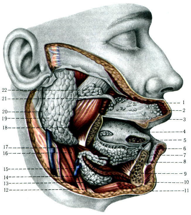
Gland ducts. Intercalary ducts are located in alveoli formed by secretory cells. The striated ducts are larger, lined with single-layer columnar epithelium and are also located inside the lobules. The union of many striated ducts forms larger interlobular ducts lined with stratified squamous epithelium.
The common excretory duct (ductus parotideus), 2-4 cm long, begins by merging all interlobular ducts, is located 1-2 cm below the zygomatic arch, on the surface masticatory muscle. At its anterior edge, it pierces the fatty body and buccal muscle, and opens into the vestibule of the mouth at the level of the second (first) large molar of the upper jaw.
The external carotid, superficial temporal, transverse, and posterior auricular arteries pass through the parotid gland. facial nerve and the retromandibular vein.
Submandibular gland
The submandibular gland (gl. submandibularis) has a lobular structure and produces a protein-mucus secretion. The gland is localized under the edge of the lower jaw in the regio submandibularis, which is limited above by m. mylohyoideus, behind - the posterior belly of the digastric muscle, in front - its anterior belly, outside - platysma. The gland is covered with a connective tissue capsule, representing part f. colli propria. General structure gland and its ducts like parotid gland. The common duct of the submandibular gland emerges on its medial surface, then penetrates between m. mylohyoideus and m. hyoglossus and reaches an elevation under the tongue - caruncula sublingualis.
Sublingual gland
The sublingual gland (gl. sublingualis) produces mucous secretion (mucin); located under the tongue and its lateral part on m. geniohyoideus. It has an alveolar structure, formed from lobules. The common duct of the gland and smaller ducts open under the tongue on the sides of the frenulum sublingualis.
The common duct often connects with the terminal part of the duct of the submandibular gland.
X-rays of the salivary glands
After introducing a contrast agent into the duct of any salivary gland (sialography), the contour and architecture of the ducts can be used to judge the condition of the gland. The contours of the duct are clear, have a uniform diameter, the architecture of the lobular ducts is correct, there are no voids; as a rule, the ducts of the 5th, 4th, 3rd, 2nd and 1st order, which have a tree-like shape, are easily filled (Fig. 225). All ducts are cleared of the contrast agent within the first hour after administration.
Embryogenesis of the salivary glands
The salivary glands develop from the epithelium of the oral cavity and grow into the surrounding mesenchyme. The parotid and submandibular glands appear in the 6th week of the intrauterine period, and the sublingual gland - in the 7th week. The terminal sections of the glands are formed from epithelium, and the connective tissue stroma, dividing the gland rudiment into lobes, is from mesenchyme.
Phylogeny of the salivary glands
Fish and aquatic amphibians do not have salivary glands. They appear only in terrestrial animals. Land amphibians acquire internasal and palatine glands. In reptiles, sublingual, labial and dental glands additionally arise. The dental glands in snakes are transformed into tubular poisonous glands located in the thickness of the masticatory muscle, and their ducts are connected to the canal or groove of the front teeth. When the masticatory muscle contracts, the venom of the gland is squeezed into the duct. Birds have sublingual glands and several small palatine glands that produce mucous saliva. Mammals have all the salivary glands, just like humans.

 Echo HSG tubal patency")


