The anatomy of the nose and paranasal sinuses has a huge clinical significance, since in close proximity to them there is not only the brain, but also many great vessels, which contribute to the rapid spread of pathogenic processes.
It is important to understand exactly how the nasal structures communicate with each other and with the surrounding space in order to understand the mechanism of development of inflammatory and infectious processes and effectively prevent them.
Nose like anatomical education, includes several structures:
- external nose;
- nasal cavity;
- paranasal sinuses.
External nose
This anatomical structure is an irregular pyramid with three sides. The external nose is very individual external signs and has a wide variety of shapes and sizes in nature.
The dorsum delimits the nose from the upper side, it ends between the eyebrows. The top of the nasal pyramid is the tip. The lateral surfaces are called wings and are clearly separated from the rest of the face by nasolabial folds. Thanks to the wings and the nasal septum, such a clinical structure as the nasal passages or nostrils is formed.
The structure of the external nose
The outer nose includes three parts
Bone frame
Its formation occurs due to the participation of the frontal and two nasal bones. The nasal bones on both sides are limited by processes coming from upper jaw. Bottom part The nasal bones participate in the formation of the pyriform opening, which is necessary for attaching the external nose.
Cartilaginous part
Lateral cartilages are necessary for the formation of the lateral nasal walls. If you go from top to bottom, you will notice the junction of the lateral cartilages with the large cartilages. The variability of the small cartilages is very high, since they are located next to the nasolabial fold and can differ in different people by quantity and form.
The nasal septum is formed by quadrangular cartilage. The clinical significance of cartilage is not only in hiding the inside of the nose, that is, organizing a cosmetic effect, but also in the fact that due to changes in the quadrangular cartilage, a diagnosis of deviated nasal septum may appear.

Soft tissues of the nose
A person does not experience a strong need for the functioning of the muscles surrounding the nose. Basically, muscles of this type perform facial functions, helping the process of identifying odors or expressing an emotional state.
The skin is closely adjacent to the tissues surrounding it, and also contains many different functional elements: glands that secrete sebum, sweat, hair follicles.
The hair that blocks the entrance to the nasal cavities performs a hygienic function, serving as additional air filters. Hair growth causes the formation of a nasal threshold.
After the nasal threshold there is a formation called the intermediate belt. It is tightly connected to the perichondral part of the nasal septum, and when deepened into the nasal cavity it transforms into the mucous membrane.
To correct a deviated nasal septum, an incision is made exactly in the place where the intermediate belt is tightly connected to the perichondrium.

Circulation
The facial and orbital arteries provide blood flow to the nose. The veins go along the way arterial vessels and are represented by the external and nasofrontal veins. The veins of the nasofrontal region merge in an anastomosis with the veins that provide blood flow to the cranial cavity. This happens due to the angular veins.
Because of this anastomosis, infection can easily spread from the nasal area into the cranial cavities.
Lymph flow is provided through the nasal lymphatic vessels, which flow into the facial ones, and those, in turn, into the submandibular ones.
The anterior ethmoidal and infraorbital nerves provide sensation to the nose, while facial nerve responsible for muscle movements.
The nasal cavity is limited by three formations. This:
- anterior third of the cranial base;
- eye sockets;
- oral cavity.
The nostrils and nasal passages in front are the limitation of the nasal cavity, and posteriorly it becomes top part throats. The transition places are called choanae. The nasal cavity is divided by the nasal septum into two approximately equal components. More often nasal septum may deviate slightly in either direction, but these changes are not significant.

Structure of the nasal cavity
Each of the two components has 4 walls.
Inner wall
It is created through the participation of the nasal septum and is divided into two sections. The ethmoid bone, or rather its plate, forms the posterosuperior section, and the vomer forms the posteroinferior section.
Outer wall
One of the complex formations. Consists of the nasal bone, the medial surface of the maxillary bone and its frontal process, the lacrimal bone adjacent posteriorly, and the ethmoid bone. The main space of the posterior part of this wall is formed by the participation of the palate bone and the main bone (mainly the internal plate belonging to the pterygoid process).
Bone part outer wall serves as a place for the attachment of three nasal conchae. The bottom, fornix and shells participate in the formation of a space called the common nasal passage. Thanks to the nasal conchae, three nasal passages are also formed - upper, middle and lower.
The nasopharyngeal passage is the end of the nasal cavity.
Superior and middle turbinates

Nasal turbinates
They are formed due to the participation of the ethmoid bone. The outgrowths of this bone also form the vesicular concha.
The clinical significance of this shell is explained by the fact that its large size can interfere with the normal process of breathing through the nose.
Naturally, breathing becomes difficult on the side where the concha is too large. Its infection must also be taken into account when inflammation develops in the cells of the ethmoid bone.
Lower sink
This is an independent bone that is attached to the crest of the maxillary bone and the palate bone.
The lower nasal passage has in its anterior third the mouth of a canal intended for the outflow of tear fluid. The turbinates are covered soft tissues
, which are very sensitive not only to the atmosphere, but also to inflammation.
The median passage of the nose has passages into most of the paranasal sinuses. The exception is the main sinus. There is also a semilunar fissure, the function of which is to provide communication between the middle meatus and the maxillary sinus.
Top wall
The perforated plate of the ethmoid bone provides the formation of the nasal arch. The holes in the plate give passage to the olfactory nerves into the cavity.

Bottom wall
Blood supply to the nose
The nasal cavity is supplied with blood by the sphenopalatine artery. The same artery gives off several branches to supply blood to the wall located behind. The anterior ethmoidal artery supplies the lateral wall of the nose with blood. The veins of the nasal cavity merge with the facial and ophthalmic veins. The ophthalmic branch has branches going to the brain, which is important in the development of infections.
The deep and superficial network of lymphatic vessels ensure the outflow of lymph from the cavity. The vessels here communicate well with the spaces of the brain, which is important for accounting for infectious diseases and the spread of inflammation.
The mucosa is innervated by the second and third branches of the trigeminal nerve.
Paranasal sinuses
The clinical significance and functional properties of the paranasal sinuses are enormous. They work in close contact with the nasal cavity. If the sinuses are exposed infectious disease or inflammation, this leads to complications on important organs located in close proximity to them.
The sinuses are literally dotted with various openings and passages, the presence of which contributes to the rapid development of pathogenic factors and aggravation of the situation in diseases.

Paranasal sinuses
Each sinus can cause infection to spread into the cranial cavity, eye damage and other complications.
Maxillary sinus
It has a pair and is located deep in the bone of the upper jaw. The sizes vary greatly, but the average is 10-12 cm.
The wall inside the sinus is the lateral wall of the nasal cavity. The sinus has an entrance to the cavity, located in the last part of the semilunar fossa. This wall is endowed with a relatively small thickness, and therefore it is often pierced in order to clarify the diagnosis or carry out therapy.
The wall of the upper part of the sinus has the smallest thickness. The posterior sections of this wall may not have a bone base at all, making do with cartilaginous tissue and many crevices bone tissue. The thickness of this wall is penetrated by the canal of the inferior orbital nerve. The infraorbital foramen opens this canal.
 The canal does not always exist, but this does not play any role, since if it is absent, then the nerve passes through the sinus mucosa. The clinical significance of this structure is that the risk of developing complications inside the skull or inside the orbit increases if pathogenic factor affects this sinus.
From below, the wall represents the sockets of the most posterior teeth. Most often, the roots of the tooth are separated from the sinus by only a small layer of soft tissue, which is common cause inflammation if you do not take care of the condition of your teeth.
Frontal sinus
It has a pair, is located deep in the forehead bone, in the center between the scales and the plates of part of the eye sockets. The sinuses can be delimited using a thin bone plate, and not always equally. It is possible that the plate may shift to one side. There may be holes in the plate that provide communication between the two sinuses.
The size of these sinuses is variable - they may be absent altogether, or they may have a huge distribution throughout the frontal scales and the base of the skull.
The wall in front is where the nerve of the eye exits. The exit is provided by the presence of a notch above the orbit. The notch cuts the entire upper part of the orbit of the eye. In this place, it is customary to perform a sinus opening and trephine puncture.

Frontal sinuses
The wall below is the smallest in thickness, which is why it is possible rapid spread infections from the sinus to the eye orbit.
The brain wall provides separation of the brain itself, namely the lobes of the forehead from the sinuses. It also represents a point of entry for infection.
The canal passing in the frontonasal region provides interaction between the frontal sinus and the nasal cavity. The anterior cells of the ethmoidal labyrinth, which have close contact with this sinus, often intercept inflammation or infection through it. Also in this connection are distributed tumor processes in both directions.
Lattice Maze
It is cells separated by thin partitions. The average number is 6-8, but it can be more or less. The cells are located in the ethmoid bone, which is symmetrical and unpaired.
The clinical significance of the ethmoidal labyrinth is explained by its close location to important organs. Also, the labyrinth may be adjacent to the deep parts that form the facial skeleton. The cells located in the back of the labyrinth are in close contact with the canal in which the nerve runs visual analyzer. Clinical diversity appears to be an option when the cells serve as the direct path of the canal.
Diseases affecting the labyrinth are accompanied by a variety of pains, varying in location and intensity. This is explained by the peculiarities of the innervation of the labyrinth, which is provided by a branch of the orbital nerve, called the nasociliary. The cribriform plate also provides passage for the nerves necessary for the functioning of the sense of smell. That is why, if there is swelling or inflammation in this area, olfactory disturbances are possible.

Lattice Maze
Main sinus
The sphenoid bone, with its body, provides the location of this sinus directly behind the ethmoid labyrinth. The choanae and the nasopharynx will be located on top.
In this sinus there is a septum that has a sagittal (vertical, dividing the object into right and left parts) location. It most often divides the sinus into two unequal lobes and does not allow them to communicate with each other.
The wall in front consists of a pair of formations: the ethmoidal and nasal. The first occurs in the region of the labyrinth cells located posteriorly. The wall is characterized by a very small thickness and, thanks to its smooth transition, almost merges with the wall below. In both parts of the sinus there are small round passages that allow the sphenoid sinus to communicate with the nasopharynx.
The wall at the back has a frontal position. How larger size sinuses, the thinner this septum is, which increases the likelihood of injury during surgical interventions in this area.
 The wall on top is the bottom region of the sella turcica, which is the location of the pituitary gland and the chiasm of the nerve that provides vision. Often, if the inflammatory process affects the main sinus, it spreads to the optic chiasm.
The wall below is the vault of the nasopharynx.
The walls on the sides of the sinus are close to the bundles of nerves and vessels that are located on the side of the sella turcica.
In general, infection of the main sinus can be called one of the most dangerous. The sinus is closely adjacent to many brain structures, for example, the pituitary gland, subarachnoid and arachnoid membranes, which makes it easier for the process to spread to the brain and can be fatal.
Pterygopalatine fossa
Located behind the tubercle of the mandibular bone. A large number of nerve fibers pass through it, so the significance of this fossa in a clinical sense is difficult to exaggerate. Inflammation of the nerves passing through this fossa is associated with a large number of symptoms in neurology.
It turns out that the nose and the formations that are closely connected with it are a very complex anatomical structure. Treatment of diseases affecting the nasal systems requires maximum care and caution from the doctor due to the close location of the brain. The main task of the patient is not to let the disease progress, bringing it to a dangerous limit, and to promptly seek help from a doctor.
There is a distinction between the external nose and the nasal cavity.
Internal structure The nose consists of a hard bony part and a soft cartilaginous part. The nasal bones are located at the top of the nose and are shaped like a pyramid. They form the base of the nose and make up the upper third of the nose. The lower two-thirds of the nose is made of cartilage. Cartilage gives shape to the lower part of the bridge of the nose and shape to the tip of the nose. There are two connected cartilaginous structures: the superior lateral cartilage and the inferior lateral cartilage (ala cartilage). The superior lateral cartilage connects the nasal bone to the inferior lateral cartilage. The inferior lateral cartilage is shaped like a curved "C" and has three regions: an outer portion (lateral crus), a middle portion (dome), and an internal portion (middle crus). It forms the wings of the nose.
The two median legs form a bridge between the nostrils called the columella.
The external nose has the shape of a pyramid and is formed by bones, cartilage, and muscles. The outside of the nose is covered with the same skin as the face. It distinguishes: the root, back, apex and wings of the nose. The root of the nose is located in the upper part of the face and is separated from the forehead by the bridge of the nose. The sides of the nose join together along the midline to form the dorsum of the nose. From below, the back of the nose passes into the apex of the nose; below, the wings of the nose limit the nostrils leading into the nasal cavity.
The external nose is an important part of the facial cosmetic ensemble. In the nasal cavity, a distinction is made between the nasal vestibule and the nasal cavity.
Vestibule of the nose covered from the inside by the skin of the external nose, which continues here through the nostrils. The skin of the vestibule contains hairs, sweat and sebaceous glands.
The vestibule passes into the nasal cavity, which is a canal passing into longitudinal direction through the bones of the facial skeleton and shaped like a prism. The bottom of the nasal cavity is the hard palate. The nasal cavity is lined with mucous membrane.
Nasal cavity The septum is divided into two halves: right and left; the septum distinguishes between bone and cartilaginous parts. Posteriorly, through the choanae, the nasal cavity communicates with the nasal part of the pharynx. Most of the nasal cavity is represented by the nasal passages, with which the paranasal sinuses (air cavities of the skull bones) communicate. Three nasal conchae (superior, middle and inferior), located on the lateral walls, increase the overall surface of the nasal cavity. Between the inward-facing surfaces of the conchae and the nasal septum there is a slit-like common nasal passage, and under the conchae there are nasal passages, which have the corresponding names: upper middle and lower. The nasolacrimal duct opens into the lower nasal passage, the posterior cells of the ethmoid bone and the sphenoid sinus open into the upper, and the middle and anterior cells of the ethmoid bone, frontal and maxillary sinuses into the middle passage.
Nasal mucosa, it can be distinguished into two parts that differ from each other in structure and function: respiratory and olfactory. The respiratory part occupies the area from the bottom of the nasal cavity to the middle of the middle turbinate. The mucous membrane of this area is covered with ciliated epithelium and contains a large number of glands that secrete mucus, in addition, there are many blood vessels in the submucosa.
The olfactory region occupies part of the nasal mucosa, covering the right and left superior turbinates, as well as part of the middle turbinates and the corresponding section of the nasal septum. In the olfactory region there are nerve cells, perceiving odorous substances from the inhaled air.
The paranasal sinuses include air cavities surrounding the nasal cavity and connected to it by openings ( excretory ducts). There are maxillary (maxillary), frontal, sphenoid and ethmoid sinuses. Their sizes vary from person to person; the maxillary sinus is considered the largest in volume (from 5 to 30 cm3). The inside of the sinuses is also lined with mucous membrane.
The maxillary sinuses are located in the body of the upper jaw, to the right and left of the nasal cavity. The roots of the teeth of the upper jaw (3-6) in some cases can protrude into the sinus, so the development of odontogenic lesions is possible in it inflammatory processes. The frontal sinuses are located in the frontal bone at the level of the brow ridges on the right and left. The ethmoid sinuses consist of individual cells and are located in the thickness of the ethmoid bone. The sphenoid sinus is located in the body of the sphenoid bone (behind the ethmoid bone) and is divided into two halves by a septum. Through special openings the sinus communicates with the nasal cavity.
The nose performs a variety of functions: respiratory, protective, resonant and olfactory.
Respiratory function is the main one. The nose is the first to perceive inhaled air, which is warmed, purified and moistened here, therefore nasal breathing is the most physiological for the body.
The protective function is that the receptors of the mucous membrane react to many stimuli from the external environment: chemical composition, temperature, humidity, dust content and other air properties. When the mucous membrane is exposed to irritants, sneezing and lacrimation appear. Tear entering the nasal cavity through the nasolacrimal duct enhances the secretion of mucous glands and removes irritants from the nasal cavity.
In the mechanical removal of substances suspended in the inhaled air important role plays the ciliated epithelium of the nasal mucosa. When the cilia vibrate, directed from the entrance to the nose to the nasopharynx, particles trapped in the nasal cavity move. Some of the larger dust particles are retained in the area of the vestibule of the nose by hairs, and if dust particles suspended in the air still enter the nasal cavity, they are removed from it with mucus when sneezing or blowing the nose. TO defense mechanisms This also includes warming and humidifying the air entering through the nose.
The resonator function is ensured by the presence of air cavities (nasal cavity, paranasal sinuses). The unequal size of these cavities contributes to the amplification of vocal tones of different frequencies. Forming in the glottis and passing through the resonator cavities, the sound acquires a certain timbre (color).
The olfactory function is carried out due to the presence of specific olfactory receptors in the nasal cavity. In human life, odors play an important role, helping to determine the good quality of food and the presence of harmful impurities in the inhaled air. In a number of cases, smell helps a person navigate the environment, experience pleasure or disgust. The sense of smell is greatly influenced by air humidity, temperature, Atmosphere pressure, general state person.
The nose of a newborn baby is flattened, short, the nasal cavity is narrow and low, poorly developed. With age, the bridge of the nose lengthens, forming the apex of the nose. During puberty, the shape of the external nose becomes constant. The paranasal sinuses in newborns are poorly developed. By the age of 8-9 years, the process of formation of the maxillary sinus ends, and by the age of 12-14 years, the sinuses of the frontal, ethmoid and sphenoid bones take their final shape.
In fact, this organ is a pair, that is, there are two nasal cavities. They are separated from each other by the nasal septum. Each nostril opens at the front, and at the back it is connected to the nasopharynx by special openings. However, it so happened that these two departments are combined in speech under the name “nasal cavity”.
Its structure is more complex than it seems to an ignorant person. The walls of the nasal cavities, the bottom and roof of the cavity are rigid due to bone, cartilage and connective tissue high density. It is because of this structural feature that the cavity does not collapse when inhaling.
Each nasal cavity is divided into two parts: the vestibule - an expanded area directly behind the nostrils, the respiratory cavity - a narrowed part located immediately behind the vestibule. The epidermis, which lines the cavity from the inside, contains a lot hair follicles, and also sweat and sebaceous glands. Why exactly is the nasal cavity lined this way? Its functions are cleansing, increasing humidity and air temperature, which is why it is so abundantly blood vessels. Hairs can trap large particles in the inhaled air.
In the vestibule, the multilayered one belongs to the non-keratinizing type, then it becomes multirowed cylindrical ciliated, and goblet cells begin to appear in it. The epithelium becomes part of the mucous membrane lining respiratory part nasal cavity.
The lamina propria of the mucous membrane here is adjacent to the periosteum or perichondrium, depending on whether this mucous membrane covers bone or cartilage. The basement membrane, which separates the respiratory epithelium from the lamina propria, is much thicker than in most other types of epithelium.
The epithelial surface is moistened with mucus, which is also produced by glands from the lamina propria. Up to 500 ml of mucus is produced per day. The latter mixes with particles of dirt and dust that stick to it, and thanks to the cilia, it moves to. Cleansing the nasal cavity largely depends on the condition of the cilia; if they have suffered from illness or injury, this process can be greatly disrupted.
In some places near the vestibule there are lymphatic follicles that perform an immune function. In the lamina propria of the nasal mucosa there are many plasma cells and lymphocytes, and sometimes granular leukocytes are also found. They “protect the borders” of the body, protecting us from invasions, because the nasal cavity often becomes the gateway to infections.
However, the cavity “works” not only with air; on the upper part of the walls, as well as the roof of the rear part of each area, there are special cells that make up the organ of smell.
There are two olfactory zones, one in each nasal cavity. The mucous membrane there forms a special organ, thanks to which we are able to smell. The peculiarity of this sensory organ is that the bodies of neurons there are located on the surface, which makes them truly vulnerable. Therefore, in case of injuries to the nose or chronic diseases a person may lose their sense of smell. We lose another approximately one percent of our sense of smell for every year of life, which is why this important sense is so often impaired in older people.
Along the side plate of each cavity there are three bone plates, one above the other, like small shelves. They are slightly curved downwards, which is why they are called turbinates.
Also associated with the nasal cavity are those found in the bone cavities. The largest is located in the smaller sinuses - in the frontal, ethmoid and sphenoid bones. They are the ones that fill with mucus and sometimes pus during sinusitis. In this case, medications are prescribed that cause the patency of the sinuses to increase.
The nasal cavity is complex, because it must protect us, prepare air for the lungs and carry out the sense of smell.
The nasal cavity is the beginning respiratory tract person. This is the air passage that connects the nasopharynx to external environment. The nasal cavity houses the olfactory organs; in addition, the incoming air is warmed and purified here.
Structure
The outer side of the nose consists of the nostrils or wings, the middle part or back and the root, which is located in the frontal lobe of the face. The bones of the skull form its walls, and the palate limits it on the side of the mouth. The entire nasal cavity is divided into two nostrils, each of which has a lateral, medial, superior, inferior and posterior wall.
The nasal cavity is built with the help of bone, membranous and cartilaginous tissue. The whole of it is divided into three shells, but only the last of them is considered true, since it is formed by bone. Between the shells there are passages through which air passes; these are the top passage, the middle passage and the bottom passage.
On inside The cavity contains the mucous membrane. The mucous membrane has a small thickness and performs several functions at once: it cleans and warms the air, and also helps to distinguish odors.
Functions
The main functions of the nasal cavity:
- respiratory function, which ensures the supply of oxygen to body tissues;
- protective function that guarantees cleaning from dust, dirt and harmful microorganisms, humidification and warming of air;
- resonator function, which guarantees the imparting of sonority and individual coloring to the voice;
- olfactory function, which allows you to distinguish different shades aromas.
Diseases of the nasal cavity
The most common diseases:
- vasomotor rhinitis, which is caused by a decrease in vascular tone from the submucosa of the lower conchae;
- allergic rhinitis, which occurs due to an individual reaction to irritants;
- hypertrophic rhinitis, which occurs as a result of other types of rhinitis and is characterized by an increase in connective tissue;
- medicinal rhinitis develops due to improper use of medications;
- adhesions after nasal injuries or surgical interventions;
- polyps, which are growths of the nasal mucosa due to advanced rhinosinusitis;
- neoplasms, which include osteomas, papillomas, fibromas, cysts.
Treatment of any diseases of the nose should be carried out immediately and professionally, since breathing problems can lead to disruptions in the functioning of almost all human organs.
Studies of the nose and paranasal sinuses
The examination of the nasal cavity is usually carried out in three stages. At the first stage, an external examination of the nose and examination of the projection sites of the paranasal sinuses on the face are carried out. The external nose, anterior and inferior walls of the frontal sinuses, anterior walls of the maxillary sinuses, submandibular and cervical lymph nodes are palpated.
At the second stage, rhinoscopy is performed, which can be anterior, middle and posterior. It is carried out using special lighting, for example, a frontal reflector or an autonomous light source. For better implementation During the examination, a nasal speculum is used - a nasal dilator. And at the last stage, the respiratory and olfactory functions of the nasal cavity are assessed.
The initial section of the upper respiratory tract consists of three parts.
Three components of the nose
- external nose
- nasal cavity
- paranasal sinuses, which communicate with the nasal cavity through narrow openings
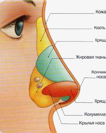
Appearance and external structure of the external nose
External nose
External nose- This is an osteochondral formation, covered with muscles and skin, in appearance resembling a hollow trihedral pyramid of irregular shape.
Nasal bones- This is the paired basis of the external nose. Attached to the nasal part of the frontal bone, they, joining each other in the middle, form the back of the external nose in its upper part.
Cartilaginous part of the nose, being a continuation of the bony skeleton, is firmly fused to the latter and forms the wings and the tip of the nose.
The wing of the nose, in addition to the larger cartilage, includes connective tissue formations from which the posterior sections of the nasal openings are formed. The inner sections of the nostrils are formed by the movable part of the nasal septum - the columella.
Muscular skin. The skin of the external nose has many sebaceous glands (mainly in the lower third of the external nose); a large number of hairs (in the vestibule of the nose), performing protective function; as well as an abundance of capillaries and nerve fibers (this explains the pain of nasal injuries). The muscles of the external nose are designed to compress the nasal openings and pull down the wings of the nose.
Nasal cavity
The entrance “gate” of the respiratory tract, through which inhaled (as well as exhaled) air passes, is the nasal cavity - the space between the anterior cranial fossa and the oral cavity.
The nasal cavity, divided by the osteochondral nasal septum into the right and left halves and communicating with the external environment through the nostrils, also has posterior openings - choanae, leading to the nasopharynx.
Each half of the nose consists of four walls. The lower wall (bottom) is the bones hard palate; the upper wall is a thin bone plate, similar to a sieve, through which branches of the olfactory nerve and vessels pass; the inner wall is the nasal septum; the lateral wall, formed by several bones, has the so-called nasal turbinates.
The turbinates (inferior, middle and superior) divide the right and left halves of the nasal cavity into tortuous nasal passages - upper, middle and lower. In the upper and middle nasal passages there are small openings through which the nasal cavity communicates with the paranasal sinuses. In the lower nasal passage there is an opening of the nasolacrimal canal, through which tears flow into the nasal cavity.
Three areas of the nasal cavity
- vestibule
- respiratory region
- olfactory region

Major bones and cartilages of the nose
Very often the nasal septum is curved (especially in men). This leads to difficulty breathing and, as a result, surgical intervention.
The vestibule limited by the wings of the nose, its edge is lined with a 4-5 mm strip of skin, equipped with a large number of hairs.
Respiratory area- this is the space from the bottom of the nasal cavity to the lower edge of the middle turbinate, lined with a mucous membrane formed by many goblet cells that secrete mucus.
U common man the nose can distinguish about ten thousand smells, and the taster can distinguish many more.
The surface layer of the mucous membrane (epithelium) has special cilia with a flickering movement directed towards the choanae. Under the mucous membrane of the nasal turbinates lies a tissue consisting of a plexus of blood vessels, which promotes instant swelling of the mucous membrane and narrowing of the nasal passages under the influence of physical, chemical and psychogenic irritants.
Nasal mucus, which has antiseptic properties, destroys great amount microbes trying to enter the body. If there are a lot of microbes, the volume of mucus also increases, which leads to a runny nose.
A runny nose is the most common disease in the world, which is why it is even included in the Guinness Book of Records. On average, an adult gets a runny nose up to ten times a year, and spends a total of up to three years with a stuffy nose throughout his life.
Olfactory region(olfactory organ), colored yellowish-brown, occupies part of the upper nasal passage and the posterosuperior part of the septum; its border is the lower edge of the middle turbinate. This zone is lined with epithelium containing olfactory receptor cells.
Olfactory cells are spindle-shaped and end on the surface of the mucous membrane with olfactory vesicles equipped with cilia. The opposite end of each olfactory cell continues into a nerve fiber. Such fibers, connecting into bundles, form the olfactory nerves (I pair). Odorous substances, entering the nose along with the air, they reach the olfactory receptors by diffusion through the mucus covering the sensitive cells, chemically interact with them and cause excitation in them. This excitation travels along the fibers of the olfactory nerve to the brain, where odors are distinguished.
When eating, the olfactory sensations complement the gustatory ones. With a runny nose, the sense of smell is dulled and food seems tasteless. With the help of smell, the smell of undesirable impurities in the atmosphere is detected; sometimes it is possible to distinguish poor-quality food from food that is suitable for eating by smell.
Olfactory receptors are very sensitive to odors. To excite the receptor, it is enough for only a few molecules of an odorous substance to act on it.

Structure of the nasal cavity
- Our smaller brothers - animals - are more partial to smells than humans.
- Birds, fish, and insects sense odors at great distances. Petrels, albatrosses, and fulmars are able to smell fish at a distance of 3 km or more. It has been confirmed that pigeons find their way by smell, flying for many kilometers.
- For moles, their hypersensitive sense of smell is a sure guide to underground labyrinths.
- Sharks smell blood in the water even at a concentration of 1:100,000,000.
- It is believed that the male moth has the most acute sense of smell.
- Butterflies almost never land on the first flower they come across: they sniff and circle over the flowerbed. Very rarely, butterflies are attracted to poisonous flowers. If this happens, the “victim” sits down by a puddle and drinks heavily.
Paranasal (paranasal) sinuses
Paranasal sinuses (sinusitis)- these are air cavities (paired), located in the front part of the skull around the nose and communicating with its cavity through the outlet openings (ostia).
Maxillary sinus- the largest (the volume of each sinus is about 30 cm 3) - located between the lower edge of the orbits and the dentition of the upper jaw.
On the inner wall of the sinus, bordering the nasal cavity, there is an anastomosis leading to the middle meatus of the nasal cavity. Since the hole is located almost under the “roof” of the sinus, this complicates the outflow of contents and contributes to the development of congestive inflammatory processes.
The anterior, or facial, wall of the sinus has a depression called the canine fossa. This area is usually where the sinus is opened during surgery.
The upper wall of the sinus is also the lower wall of the orbit. Bottom maxillary sinus very close to the roots of the rear upper teeth, to the point that sometimes the sinus and teeth are separated only by the mucous membrane, and this can lead to sinus infection.
The maxillary sinus got its name from the English doctor Nathaniel Highmore, who first described its diseases

Diagram of the location of the paranasal sinuses
Fat back wall The sinuses are bordered by the cells of the ethmoidal labyrinth and the sphenoid sinus.
Frontal sinus is located in the thickness of the frontal bone and has four walls. Using a thin winding canal that opens into the anterior section of the middle meatus, the frontal sinus communicates with the nasal cavity. Bottom wall The frontal sinus is the upper wall of the orbit. The median wall separates the left frontal sinus from the right, the posterior wall separates the frontal sinus from the frontal lobe of the brain.
Ethmoid sinus, also called the “labyrinth,” is located between the orbit and the nasal cavity and consists of individual air-bearing bony cells. There are three groups of cells: anterior and middle, opening into the middle nasal meatus, and posterior, opening into the upper nasal meatus.
Sphenoid (main) sinus lies deep in the body of the sphenoid (main) bone of the skull, divided by a septum into two separate halves, each of which has an independent exit to the area of the upper nasal passage.
At birth, a person has only two sinuses: the maxillary and the ethmoidal labyrinth. The frontal and sphenoid sinuses are absent in newborns and begin to form only from 3-4 years of age. The final development of the sinuses ends at approximately 25 years of age.
Functions of the nose and paranasal sinuses
The complex structure of the nose ensures that it successfully performs the four functions assigned to it by nature.
Olfactory function. The nose is one of the most important sense organs. With its help, a person perceives all the variety of smells around him. The loss of smell not only impoverishes the palette of sensations, but is also fraught negative consequences. After all, some odors (for example, the smell of gas or spoiled food) signal danger.
Respiratory function- most important. It ensures the supply of oxygen to the body tissues, which is necessary for normal functioning and blood gas exchange. When nasal breathing is difficult, the course of oxidative processes in the body changes, which leads to disruption of the cardiovascular and nervous systems, disorders of the lower respiratory tract and gastrointestinal tract, increased intracranial pressure.
The aesthetic significance of the nose plays an important role. Often, while ensuring normal nasal breathing and sense of smell, the shape of the nose gives its owner significant experiences, not corresponding to his ideas of beauty. In this regard, it is necessary to resort to plastic surgery, corrective appearance external nose.
Protective function. The inhaled air, passing through the nasal cavity, is cleared of dust particles. Large dust particles are trapped by hairs that grow at the entrance to the nose; Some of the dust particles and bacteria, passing along with the air into the winding nasal passages, settle on the mucous membrane. Non-stop vibrations of the cilia of the ciliated epithelium remove mucus from the nasal cavity into the nasopharynx, from where it is expectorated or swallowed. Bacteria that enter the nasal cavity are largely neutralized by substances contained in nasal mucus. Cold air, passing through the narrow and winding nasal passages, is warmed and moistened by the mucous membrane, which is abundantly supplied with blood.
Resonator function. The nasal cavity and paranasal sinuses can be compared to sound system: the sound, reaching their walls, intensifies. The nose and sinuses play a leading role in the pronunciation of nasal consonants. Nasal congestion causes nasal sounds, in which nasal sounds are pronounced incorrectly.


