Trillions of cells in human body found in all shapes and sizes. These tiny structures are the core. Cells form organ tissues, which form organ systems that work together to maintain the body's functioning.
There are hundreds of different types of cells in the body, and each type is suited to the role it serves. Cells digestive system, for example, differ in structure and function from cells of the skeletal system. Regardless of differences, the cells of the body depend on each other, directly or indirectly, in order for the body to function as a whole. Below are examples of the different types of cells in the human body.
Stem cells
Stem cells are unique cells in the body because they are unspecialized and have the ability to develop into specialized cells for specific organs or tissues. Stem cells are capable of dividing multiple times to replenish and repair tissue. In the field of stem cell research, scientists are trying to take advantage of the renewable properties by using them to create cells for tissue repair, organ transplantation and disease treatment.
Bone cells
Bones are a type of mineralized connective tissue and a major component of the skeletal system. Bone cells form bone, which is composed of a matrix of the minerals collagen and calcium phosphate. There are three main types of bone cells in the body. Osteoclasts are large cells that break down bone for resorption and assimilation. Osteoblasts regulate bone mineralization and produce osteoid (organic bone matrix matter). Osteoblasts mature to form osteocytes. Osteocytes help in bone formation and maintain calcium balance.
Blood cells
From transporting oxygen throughout the body to fighting infection, cells are vital to life. There are three main types of cells in the blood - red blood cells, white blood cells and platelets. Red blood cells determine the type of blood and are also responsible for transporting oxygen to the cells. Leukocytes are cells immune system, which destroy and provide immunity. Platelets help thicken the blood and prevent excessive blood loss from damaged blood vessels. Blood cells are produced by the bone marrow.
Muscle cells
Muscle cells form muscle tissue, which is important for bodily movement. Skeletal muscle Attaches to bones to aid movement. Skeletal muscle cells covered with connective tissue that protects and supports muscle fiber bundles. Cardiac muscle cells form the involuntary heart muscle. These cells assist in the contraction of the heart and are connected to each other through intercalated discs to synchronize the heart rhythm. Smooth muscle tissue is not stratified like cardiac or skeletal muscle. Smooth muscle is an involuntary muscle that forms body cavities and the walls of many organs (kidneys, intestines, blood vessels, respiratory tract lungs, etc.).
Fat cells
Fat cells, also called adipocytes, are the main cellular component of adipose tissue. Adipocytes contain triglycerides, which can be used for energy. During fat storage, fat cells swell and become round in shape. When fat is used, these cells decrease in size. Fat cells also have an endocrine function, since they produce hormones that affect the metabolism of sex hormones, regulation blood pressure, insulin sensitivity, fat storage or utilization, blood clotting, and cell signaling.
Skin cells
The skin is made up of a layer epithelial tissue(epidermis), which is supported by a layer of connective tissue (dermis) and subcutaneous layer. The outermost layer of skin is made up of flat epithelial cells, which are tightly packed together. Skin protects internal structures the body from damage, prevents dehydration, acts as a barrier against microbes, stores fat, produces vitamins and hormones.
Nerve cells (neurons)
Nerve tissue cells or neurons are the basic unit nervous system. Nerves transmit signals between the brain spinal cord and organs of the body through nerve impulses. A neuron consists of two main parts: the cell body and neural processes. The central cell body includes neural, associated and. Nervous processes are "finger-like" projections (axons and dendrites) extending from the cell body and are capable of conducting or transmitting signals.
Endothelial cells
Endothelial cells form the inner lining of cardio-vascular system and structures of the lymphatic systems. These cells make up the inner layer of blood vessels, lymphatic vessels and organs, including the brain, lungs, skin and heart. Endothelial cells are responsible for angiogenesis, or the creation of new blood vessels. They also regulate the movement of macromolecules, gases and fluids between the blood and surrounding tissues, and help regulate blood pressure.
Sex cells
Cancer cells
Cancer is the result of the development of abnormal properties in normal cells, which allows them to divide uncontrollably and spread elsewhere in the body. Development can be caused by mutations that occur from factors such as chemicals, radiation, ultraviolet radiation, replication errors, or viral infection. Cancer cells lose sensitivity to anti-growth signals, multiply rapidly and lose the ability to undergo .
All cellular life forms on earth can be divided into two superkingdoms based on the structure of their constituent cells - prokaryotes (prenuclear) and eukaryotes (nuclear). Prokaryotic cells are simpler in structure; apparently, they arose earlier in the process of evolution. Eukaryotic cells are more complex and arose later. The cells that make up the human body are eukaryotic.
Despite the variety of forms, the organization of cells of all living organisms is subject to common structural principles.
Prokaryotic cell
Eukaryotic cell
Structure of a eukaryotic cell
Superficial complex animal cell
Comprises glycocalyx, plasma membranes and the cortical layer of cytoplasm located underneath. The plasma membrane is also called plasmalemma, the outer cell membrane. This is a biological membrane, about 10 nanometers thick. Provides primarily a delimiting function in relation to the environment external to the cell. In addition, it performs a transport function. The cell does not waste energy to maintain the integrity of its membrane: the molecules are held together according to the same principle by which fat molecules are held together - it is thermodynamically more advantageous for the hydrophobic parts of the molecules to be located in close proximity to each other. The glycocalyx is molecules of oligosaccharides, polysaccharides, glycoproteins and glycolipids “anchored” in the plasmalemma. The glycocalyx performs receptor and marker functions. The plasma membrane of animal cells mainly consists of phospholipids and lipoproteins interspersed with protein molecules, in particular surface antigens and receptors. In the cortical (adjacent to the plasma membrane) layer of the cytoplasm there are specific cytoskeletal elements - actin microfilaments ordered in a certain way. The main and most important function of the cortical layer (cortex) is pseudopodial reactions: ejection, attachment and contraction of pseudopodia. In this case, the microfilaments are rearranged, lengthened or shortened. The shape of the cell (for example, the presence of microvilli) also depends on the structure of the cytoskeleton of the cortical layer.
Cytoplasmic structure
The liquid component of the cytoplasm is also called cytosol. Under a light microscope, it seemed that the cell was filled with something like liquid plasma or sol, in which the nucleus and other organelles “floated”. Actually this is not true. The internal space of a eukaryotic cell is strictly ordered. The movement of organelles is coordinated with the help of specialized transport systems, the so-called microtubules, which serve as intracellular “roads” and special proteins dyneins and kinesins, which play the role of “motors”. Individual protein molecules also do not diffuse freely throughout the entire intracellular space, but are directed to the necessary compartments using special signals on their surface, recognized by the cell’s transport systems.
Endoplasmic reticulum
In a eukaryotic cell, there is a system of membrane compartments (tubes and cisterns) passing into each other, which is called the endoplasmic reticulum (or endoplasmic reticulum, ER or EPS). That part of the ER, to the membranes of which ribosomes are attached, is referred to as granular(or rough) endoplasmic reticulum, protein synthesis occurs on its membranes. Those compartments that do not have ribosomes on their walls are classified as smooth(or agranular) ER, which takes part in lipid synthesis. The internal spaces of the smooth and granular ER are not isolated, but pass into each other and communicate with the lumen of the nuclear envelope.
Golgi apparatus
Core
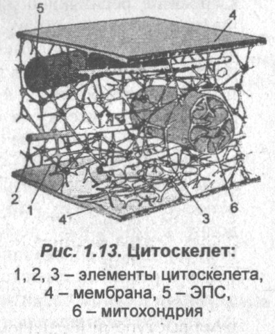
Cytoskeleton
Centrioles
Mitochondria
Comparison of pro- and eukaryotic cells
The most important difference between eukaryotes and prokaryotes for a long time the presence of a formed core was considered and membrane organelles. However, by the 1970-1980s. it became clear that this was only a consequence of deeper differences in the organization of the cytoskeleton. For some time it was believed that the cytoskeleton is characteristic only of eukaryotes, but in the mid-1990s. proteins homologous to the main proteins of the cytoskeleton of eukaryotes have also been discovered in bacteria.
It is the presence of a specifically structured cytoskeleton that allows eukaryotes to create a system of mobile internal membrane organelles. In addition, the cytoskeleton allows endo- and exocytosis to occur (it is assumed that it was thanks to endocytosis that intracellular symbionts, including mitochondria and plastids, appeared in eukaryotic cells). Other most important function cytoskeleton of eukaryotes - ensuring the division of the nucleus (mitosis and meiosis) and body (cytotomy) of a eukaryotic cell (the division of prokaryotic cells is organized more simply). Differences in the structure of the cytoskeleton also explain other differences between pro- and eukaryotes - for example, the constancy and simplicity of the forms of prokaryotic cells and the significant diversity of shape and the ability to change it in eukaryotic cells, as well as the relatively large size of the latter. Thus, the sizes of prokaryotic cells average 0.5-5 microns, the sizes of eukaryotic cells average from 10 to 50 microns. In addition, only among eukaryotes are there truly giant cells, such as the massive eggs of sharks or ostriches (in a bird egg, the entire yolk is one huge egg), neurons large mammals, the processes of which, strengthened by the cytoskeleton, can reach tens of centimeters in length.
Anaplasia
The destruction of cellular structure (for example, in malignant tumors) is called anaplasia.
History of cell discovery
The first person to see cells was the English scientist Robert Hooke (known to us thanks to Hooke's law). In the year, trying to understand why the cork tree floats so well, Hooke began to examine thin sections of cork using a microscope he had improved. He discovered that the cork was divided into many tiny cells, which reminded him of monastery cells, and he called these cells cells (in English cell means “cell, cell, cell”). In the same year, the Dutch master Anton van Leeuwenhoek (-) used a microscope for the first time to see “animals” - moving living organisms - in a drop of water. Thus, by the beginning of the 18th century, scientists knew that under high magnification plants have a cellular structure, and they saw some organisms that were later called unicellular. However, the cellular theory of the structure of organisms was formed only in the middle of the 19th century, after more powerful microscopes appeared and methods for fixing and staining cells were developed. One of its founders was Rudolf Virchow, but his ideas contained a number of errors: for example, he assumed that cells were weakly connected to each other and each existed “on its own.” Only later was it possible to prove the integrity of the cellular system.
see also
- Comparison of the cell structure of bacteria, plants and animals
Links
- Molecular Biology Of The Cell, 4th edition, 2002 - textbook on molecular biology in English
- Cytology and Genetics (0564-3783) publishes articles in Russian, Ukrainian and English at the author’s choice, translated into English language (0095-4527)
The cells that make up the tissues of plants and animals vary significantly in shape, size and internal structure. However, they all show similarities in the main features of life processes, metabolism, irritability, growth, development, and the ability to change.
Biological transformations occurring in a cell are inextricably linked with those structures of a living cell that are responsible for performing one or another function. Such structures are called organelles.
Cells of all types contain three main, inextricably linked components:
- structures that form its surface: the outer membrane of the cell, or cell wall, or cyto plasma membrane;
- cytoplasm with a whole complex of specialized structures - organelles (endoplasmic reticulum, ribosomes, mitochondria and plastids, Golgi complex and lysosomes, the cell center), permanently present in the cell, and temporary formations called inclusions;
- nucleus - separated from the cytoplasm by a porous membrane and contains nuclear sap, chromatin and nucleolus.
Cell structure
The surface apparatus of the cell (cytoplasmic membrane) of plants and animals has some features.
In unicellular organisms and leukocytes, the outer membrane ensures the penetration of ions, water, and small molecules of other substances into the cell. The process of penetration of solid particles into a cell is called phagocytosis, and the entry of droplets of liquid substances is called pinocytosis.
The outer plasma membrane regulates the exchange of substances between the cell and the external environment.
Eukaryotic cells contain organelles covered with a double membrane - mitochondria and plastids. They contain their own DNA and protein-synthesizing apparatus, reproduce by division, that is, they have a certain autonomy in the cell. In addition to ATP, small amounts of protein are synthesized in mitochondria. Plastids are characteristic of plant cells and reproduce by division.
| Types of cells | Structure and functions of external and inner layers cell membrane | ||
|---|---|---|---|
| outer layer (chemical composition, functions) |
inner layer - plasma membrane |
||
| chemical composition | functions | ||
| Plant cells | Consist of fiber. This layer serves as the frame of the cell and performs a protective function. | Two layers of protein, between them is a layer of lipids | Limits the internal environment of the cell from the external and maintains these differences |
| Animal cells | The outer layer (glycocalyx) is very thin and elastic. Consists of polysaccharides and proteins. Performs a protective function. | Same | Special enzymes of the plasma membrane regulate the penetration of many ions and molecules into the cell and their release into the external environment |
Single-membrane organelles include the endoplasmic reticulum, Golgi complex, lysosomes, and various types of vacuoles.
Modern research tools have allowed biologists to establish that, according to the structure of the cell, all living beings should be divided into “non-nuclear” organisms - prokaryotes and “nuclear” - eukaryotes.
Prokaryotes-bacteria and blue-green algae, as well as viruses, have only one chromosome, represented by a DNA molecule (less commonly RNA), located directly in the cytoplasm of the cell.
| Main organoids | Structure | Functions |
|---|---|---|
| Cytoplasm | Internal semi-liquid medium of fine-grained structure. Contains nucleus and organelles |
|
| ER - endoplasmic reticulum | A membrane system in the cytoplasm" that forms channels and larger cavities; EPS is of 2 types: granular (rough), on which many ribosomes are located, and smooth |
|
| Ribosomes | Small bodies with a diameter of 15-20 mm | Carry out the synthesis of protein molecules and their assembly from amino acids |
| Mitochondria | They have spherical, thread-like, oval and other shapes. Inside the mitochondria there are folds (length from 0.2 to 0.7 µm). The outer cover of mitochondria consists of 2 membranes: the outer one is smooth, and the inner one forms cross-shaped outgrowths on which respiratory enzymes are located |
|
| Plastids are characteristic only of plant cells and come in three types: | Double-membrane cell organelles | |
| chloroplasts | Have green color, oval in shape, limited from the cytoplasm by two three-layer membranes. Inside the chloroplast there are edges where all the chlorophyll is concentrated | Use light energy from the sun and create organic substances from inorganic ones |
| chromoplasts | Yellow, orange, red or brown, formed as a result of the accumulation of carotene | Give various parts plants red and yellow colors |
| leucoplasts | Colorless plastids (found in roots, tubers, bulbs) | They store reserve nutrients |
| Golgi complex | May have different shapes and consists of cavities delimited by membranes and tubes extending from them with bubbles at the end |
|
| Lysosomes | Round bodies with a diameter of about 1 micron. They have a membrane (skin) on the surface, inside which there is a complex of enzymes | Perform a digestive function - digest food particles and remove dead organelles |
| Cell movement organelles |
|
|
| Cellular inclusions | These are the unstable components of the cell - carbohydrates, fats and proteins | Spare nutrients used during cell life |
| Cell center | Consists of two small bodies - centrioles and centrosphere - a compacted section of the cytoplasm | Playing important role during cell division |
Eukaryotes have a great wealth of organelles and have nuclei containing chromosomes in the form of nucleoproteins (a complex of DNA with the protein histone). Eukaryotes include most modern plants and animals, both unicellular and multicellular.
There are two levels of cellular organization:
- prokaryotic - their organisms are very simply structured - these are unicellular or colonial forms that make up the kingdom of shotguns, blue-green algae and viruses
- eukaryotic - unicellular colonial and multicellular forms, from the simplest - rhizomes, flagellates, ciliates - to higher plants and animals, constituting the plant kingdom, the fungal kingdom, the animal kingdom

| Major organelles | Structure | Functions |
|---|---|---|
| Nucleus of plant and animal cells | Round or oval shape | |
| The nuclear envelope consists of 2 membranes with pores |
|
|
| Nuclear juice (karyoplasm) - semi-liquid substance | Environment in which nucleoli and chromosomes are located | |
| Nucleoli are spherical or irregular in shape | They synthesize RNA, which is part of the ribosome | |
| Chromosomes are dense, elongated or thread-like structures visible only during cell division | Contain DNA, which contains hereditary information that is passed on from generation to generation |
All cell organelles, despite the peculiarities of their structure and functions, are interconnected and “work” for the cell as a single system in which the cytoplasm is the connecting link.
Special biological objects that occupy an intermediate position between living and inanimate nature are viruses, discovered in 1892 by D.I. Ivanovsky; they currently constitute the object of a special science - virology.
Viruses reproduce only in the cells of plants, animals and humans, causing various diseases. Viruses have a very layered structure and consist of nucleic acid (DNA or RNA) and a protein shell. Outside the host cells, the viral particle does not exhibit any vital functions: it does not feed, does not breathe, does not grow, does not reproduce.
Cell is the smallest and most basic structural unit living organisms capable of self-renewal, self-regulation and self-reproduction.
Characteristic cell sizes: bacterial cells - from 0.1 to 15 microns, cells of other organisms - from 1 to 100 microns, sometimes reaching 1-10 mm; eggs of large birds - up to 10-20 cm, processes of nerve cells - up to 1 m.
Cell shape very diverse: there are spherical cells (cocci), chain (streptococci), elongated (rods or bacilli), curved (vibrios), crimped (spirilla), multifaceted, with motor flagella, etc.
Types of cells: prokaryotic(non-nuclear) and eukaryotic (having a formed nucleus).
Eukaryotic cells, in turn, are divided into cells animals, plants and fungi.
Structural organization of a eukaryotic cell
Protoplast- this is all the living contents of the cell. The protoplast of all eukaryotic cells consists of a cytoplasm (with all organelles) and a nucleus.
Cytoplasm- this is the internal contents of the cell, with the exception of the nucleus, consisting of hyaloplasm, organelles immersed in it and (in some types of cells) intracellular inclusions (reserve nutrients and/or end products of metabolism).
Hyaloplasma- main plasma, cytoplasmic matrix, the main substance, which is the internal environment of the cell and is a viscous colorless colloidal solution (water content up to 85%) of various substances: proteins (10%), sugars, organic and inorganic acids, amino acids, polysaccharides, RNA, lipids, mineral salts, etc.
■ Hyaloplasm is a medium for intracellular metabolic reactions and a connecting link between cell organelles; it is capable of reversible transitions from sol to gel; its composition determines the buffering and osmotic properties of the cell. The cytoplasm contains a cytoskeleton consisting of microtubules and contractile protein filaments.
■ The cytoskeleton determines the shape of the cell and is involved in the intracellular movement of organelles and individual substances. The nucleus is the largest organelle of a eukaryotic cell, containing chromosomes in which all hereditary information is stored (see below for more details).
Structural components of a eukaryotic cell:
■ plasmalemma (plasma membrane),
■ cell wall (only in plant cells and mushrooms),
■ biological (elementary) membranes,
■ core,
■ endoplasmic reticulum (endoplasmic reticulum),
■ mitochondria,
■ Golgi complex,
■ chloroplasts (only in plant cells),
■ lysosomes, s
■ ribosomes,
■ cell center,
■ vacuoles (only in plant and fungal cells),
■ microtubules,
■ cilia, flagella.
Schemes of the structure of animal and plant cells are given below:

Biological (elementary) membranes- These are active molecular complexes that separate intracellular organelles and cells. All membranes have a similar structure.
Structure and composition of membranes: thickness 6-10 nm; consist mainly of protein molecules and phospholipids.
■Phospholipids form a double (bimolecular) layer in which their molecules face their hydrophilic (water-soluble) ends outward and their hydrophobic (water-insoluble) ends inward of the membrane.
■ Protein molecules located on both surfaces of the lipid bilayer ( peripheral proteins), penetrate both layers of lipid molecules ( integral proteins, most of which are enzymes) or only one layer of them (semi-integral proteins).
Membrane properties: plasticity, asymmetry(the composition of the outer and inner layers of both lipids and proteins is different), polarity (the outer layer is positively charged, the inner one is negatively charged), the ability to self-close, selective permeability (in this case, hydrophobic substances pass through the lipid bilayer, and hydrophilic ones pass through the pores in integral proteins ).
Membrane functions: barrier (separates the contents of an organoid or cell from the environment), structural (provides a certain shape, size and stability of the organoid or cell), transport (ensures the transport of substances into and out of the organoid or cell), catalytic (ensures near-membrane biochemical processes), regulatory ( participates in the regulation of metabolism and energy between the organelle or cell and the external environment), participates in the conversion of energy and the maintenance of transmembrane electrical potential.
Plasma membrane (plasmalemma)
Plasma membrane, or plasmalemma, is a biological membrane or a complex of biological membranes tightly adjacent to each other, covering the cell from the outside.
The structure, properties and functions of the plasmalemma are basically the same as those of elementary biological membranes.
❖ Structural features:

■ the outer surface of the plasmalemma contains a glycocalyx - a polysaccharide layer of molecules of glycolipoids and glycoproteins that serve as receptors for “recognition” of certain chemical substances; in animal cells it may be covered with mucus or chitin, and in plant cells— cellulose or pectin substances;
■ usually the plasmalemma forms outgrowths, invaginations, folds, microvilli, etc., increasing the surface of the cell.
■ Additional functions: receptor (participates in the “recognition” of substances and in the perception of signals from the environment and transmitting them to the cell), ensuring communication between cells in tissues multicellular organism, participation in the construction of special cell structures (flagella, cilia, etc.).
Cell wall (envelope)
Cell wall is a rigid structure located outside the plasmalemma and representing the outer cover of the cell. Present in prokaryotic cells and cells of fungi and plants.
❖Cell wall composition: cellulose in plant cells and chitin in fungal cells (structural components), proteins, pectins (which are involved in the formation of plates that hold together the walls of two neighboring cells), lignin (which holds cellulose fibers together into a very strong frame), suberin (deposited on the shell from the inside and makes it is practically impermeable to water and solutions), etc. Outside surface The cell wall of plant epidermal cells contains a large amount of calcium carbonate and silica (mineralization) and is covered with hydrophobic substances waxes and cuticle (a layer of cutin substance penetrated by cellulose and pectins).
❖ Functions of the cell wall: serves as an external frame, maintains cell turgor, performs protective and transport functions.
Cell organelles
Organelles (or organelles)- These are permanent, highly specialized intracellular structures that have a specific structure and perform corresponding functions.
❖ By purpose
organelles are divided into:
■ general purpose organelles (mitochondria, Golgi complex, endoplasmic reticulum, ribosomes, centrioles, lysosomes, plastids) and
■ organelles special purpose(myofibrils, flagella, cilia, vacuoles).
❖ By the presence of a membrane
organelles are divided into:
■ double-membrane (mitochondria, plastids, cell nucleus),
■ single-membrane (endoplasmic reticulum, Golgi complex, lysosomes, vacuoles) and
■ non-membrane (ribosomes, cell center).
The internal contents of membrane organelles always differ from the hyaloplasm surrounding them.
Mitochondria- double-membrane organelles of eukaryotic cells that carry out oxidation organic matter to final products with the release of energy stored in ATP molecules.

■ Structure: rod-shaped, spherical and thread-like shapes, thickness 0.5-1 µm, length 2-7 µm; double-membrane, the outer membrane is smooth and has high permeability, the inner membrane forms folds - cristae, on which there are spherical bodies - ATP-somes. Hydrogen ions 11, which are involved in oxygen respiration, accumulate in the space between the membranes.
■ Internal contents (matrix): ribosomes, circular DNA, RNA, amino acids, proteins, Krebs cycle enzymes, tissue respiration enzymes (located on the cristae).
■ Functions: oxidation of substances to CO 2 and H 2 O; synthesis of ATP and specific proteins; the formation of new mitochondria as a result of fission in two.
Plastids(available only in plant cells and autotrophic protists).

■ Types of plastids: chloroplasts (green), leucoplasts (colorless, round in shape), chromoplasts (yellow or orange); plastids can change from one type to another.
■Structure of chloroplasts: they are double-membrane, have a round or oval shape, length 4-12 µm, thickness 1-4 µm. The outer membrane is smooth, the inner membrane has thylakoids - folds forming closed disc-shaped invaginations, between which there is stroma (see below). In higher plants, thylakoids are collected in stacks (like a column of coins) grains , which are connected to each other lamellae (single membranes).
■ Chloroplast composition: in the membranes of thylakoids and granules there are grains of chlorophyll and other pigments; internal contents (stroma): proteins, lipids, ribosomes, circular DNA, RNA, enzymes involved in CO 2 fixation, storage substances.
■Functions of plastids: photosynthesis (chloroplasts contained in the green organs of plants), synthesis of specific proteins and accumulation of reserve nutrients: starch, proteins, fats (leukoplasts), imparting color to plant tissues in order to attract pollinating insects and distributors of fruits and seeds (chromoplasts).
Endoplasmic reticulum (EPS), or endoplasmic reticulum, found in all eukaryotic cells.
■Structure: is a system of interconnected tubules, tubes, cisterns and cavities various shapes and sizes, the walls of which are formed by elementary (single) biological membranes. There are two types of EPS: granular (or rough), containing ribosomes on the surface of channels and cavities, and agranular (or smooth), not containing ribosomes.
■Functions: division of the cell cytoplasm into compartments that prevent the mixing of the chemical processes occurring in them; rough ER accumulates, isolates for maturation and transports proteins synthesized by ribosomes on its surface, synthesizes cell membranes; smooth EPS synthesizes and transports lipids, complex carbohydrates and steroid hormones, removes toxic substances from the cell.
Golgi complex (or apparatus) — membrane organelle eukaryotic cell, located near the cell nucleus, which is a system of tanks and vesicles and is involved in the accumulation, storage and transportation of substances, the construction of the cell membrane and the formation of lysosomes.

■Structure: the complex is a dictyosome - a stack of membrane-bound flat disc-shaped sacs (cisterns), from which vesicles bud, and a system of membrane tubules connecting the complex with the channels and cavities of the smooth ER.
■Functions: the formation of lysosomes, vacuoles, plasmalemma and the cell wall of a plant cell (after its division), the secretion of a number of complex organic substances (pectin substances, cellulose, etc. in plants; glycoproteins, glycolipids, collagen, milk proteins, bile, a number of hormones, etc. animals); accumulation and dehydration of lipids transported along the EPS (from smooth EPS), modification and accumulation of proteins (from granular EPS and free ribosomes of the cytoplasm) and carbohydrates, removal of substances from the cell.
Mature dictyosome cisternae lacing vesicles (Golgi vacuoles), filled with secretion, which is then either used by the cell itself or removed beyond its boundaries.
❖ Lysosomes- cellular organelles that ensure the breakdown of complex molecules of organic substances; are formed from vesicles separated from the Golgi complex or smooth ER and are present in all eukaryotic cells.
■ Structure and composition: lysosomes are small single-membrane round vesicles with a diameter of 0.2-2 µm; filled with hydrolytic (digestive) enzymes (~40), capable of breaking down proteins (to amino acids), lipids (to glycerol and higher carboxylic acids), polysaccharides (to monosaccharides) and nucleic acids (to nucleotides).
Merging with endocytic vesicles, lysosomes form a digestive vacuole (or secondary lysosome), where the breakdown of complex organic substances occurs; the resulting monomers enter the cell cytoplasm through the membrane of the secondary lysosome, and undigested (non-hydrolyzed) substances remain in the secondary lysosome and then, as a rule, are excreted outside the cell.
■ Functions: heterophagy- breakdown of foreign substances that enter the cell through endocytosis, autophagy - destruction of structures unnecessary for the cell; autolysis is the self-destruction of a cell that occurs as a result of the release of the contents of lysosomes during cell death or degeneration.
❖ Vacuoles- large vesicles or cavities in the cytoplasm that form in the cells of plants, fungi and many protists and bounded by an elementary membrane - the tonoplast.

■ Vacuoles protists are divided into digestive and contractile (having bundles of elastic fibers in the membranes and serving for osmotic regulation of the cell’s water balance).
■Vacuoles plant cells filled with cell sap - aqueous solution various organic and inorganic substances. They may also contain toxic and tannin substances and end products of cell activity.
■Vacuoles of plant cells can merge into a central vacuole, which occupies up to 70-90% of the cell volume and can be penetrated by strands of cytoplasm.
Functions: accumulation and isolation of reserve substances and substances intended for excretion; maintaining turgor pressure; ensuring cell growth due to stretching; regulation of cell water balance.
♦Ribosomes- cell organelles, present in all cells (in the amount of several tens of thousands), located on the membranes of granular EPS, in mitochondria, chloroplasts, cytoplasm and outer nuclear membrane and carrying out the biosynthesis of proteins; Ribosomal subunits are formed in the nucleoli.

■ Structure and composition: ribosomes are the smallest (15-35 nm) non-membrane granules of round and mushroom shape; have two active centers (aminoacyl and peptidyl); consist of two unequal subunits - a large one (in the form of a hemisphere with three protrusions and a channel), which contains three RNA molecules and a protein, and a small one (containing one RNA molecule and a protein); the subunits are connected using the Mg+ ion.
■ Function: synthesis of proteins from amino acids.
❖ Cell center- an organelle of most animal cells, some fungi, algae, mosses and ferns, located (in interphase) in the center of the cell near the nucleus and serving as the assembly initiation center microtubules .
■ Structure: The cell center consists of two centrioles and a centrosphere. Each centriole (Fig. 1.12) looks like a cylinder 0.3-0.5 µm long and 0.15 µm in diameter, the walls of which are formed by nine triplets of microtubules, and the middle is filled with a homogeneous substance. The centrioles are located perpendicular to each other and are surrounded by a dense layer of cytoplasm with radiating microtubules forming a radiating centrosphere. During cell division, the centrioles move toward the poles.

■ Main functions: formation of cell division poles and achromatic filaments of the division spindle (or mitotic spindle), ensuring equal distribution of genetic material between daughter cells; in interphase, it directs the movement of organelles in the cytoplasm.
Cytosklst cells is a system microfilaments And microtubules , penetrating the cytoplasm of the cell, associated with the outer cytoplasmic membrane and nuclear envelope and maintaining the shape of the cell.
■Microflanges- thin, contractile filaments 5-10 nm thick and consisting of proteins ( actin, myosin and etc.). Found in the cytoplasm of all cells and pseudopods of motile cells.
Functions: microfilaments provide the motor activity of hyaloplasm, are directly involved in changing the shape of the cell during the spreading and amoeboid movement of protist cells, and participate in the formation of the constriction during the division of animal cells; one of the main elements of the cell cytoskeleton.
■ Microtubules- thin hollow cylinders (25 nm in diameter), consisting of tubulin protein molecules, arranged in spiral or straight rows in the cytoplasm of eukaryotic cells.
Functions: microtubules form spindle filaments, are part of centrioles, cilia, flagella, and participate in intracellular transport; one of the main elements of the cell cytoskeleton.
Organelles of movement — flagella and cilia , are present in many cells, but are more common in single-celled organisms.
■ Cilia- numerous cytoplasmic short (5-20 µm long) projections on the surface of the plasmalemma. Available on the surface various types animal cells and some plants.
■ Flagella- single cytoplasmic projections on the surface of the cells of many protists, zoospores and spermatozoa; ~10 times longer than cilia; are used for movement.
■ Structure: cilia and flagella (Fig. 1.14) consist of them microtubules, arranged according to the 9 × 2 + 2 system (nine double microtubules - doublets form a wall, in the middle there are two single microtubules). The doublets are able to slide past each other, which leads to the bending of the cilium or flagellum. At the base of flagella and cilia there are basal bodies, identical in structure to centrioles.

■ Functions: cilia and flagella ensure the movement of the cells themselves or the surrounding fluid and particles suspended in it.
Inclusions
Inclusions- non-permanent (temporarily existing) components of the cell cytoplasm, the content of which varies depending on the functional state of the cell. There are trophic, secretory and excretory inclusions.
■ Trophic inclusions - these are reserves of nutrients (fat, starch and protein grains, glycogen).
■ Secretory inclusions- these are waste products of the endocrine and exocrine glands (hormones, enzymes).
■ Excretory inclusions- These are metabolic products in the cell that must be excreted from the cell.
Nucleus and chromosomes
Core- the largest organelle; is an obligatory component of all eukaryotic cells (with the exception of phloem sieve tube cells of higher plants and mature erythrocytes of mammals). Most cells have a single nucleus, but there are bi- and multinucleated cells. There are two states of the nucleus: interphase and fissile
Interphase nucleus comprises nuclear envelope(separating the internal contents of the nucleus from the cytoplasm), nuclear matrix (karyoplasm), chromatin and nucleoli. The shape and size of the nucleus depend on the type of organism, type, age and functional state of the cell. It has a high content of DNA (15-30%) and RNA (12%).
■ Kernel functions: storage and transmission of hereditary information in the form of an unchanged DNA structure; regulation (through the protein synthesis system) of all cell life processes.

❖ Nuclear envelope(or karyolemma) consists of outer and inner biological membranes, between which there is perinuclear space. The inner membrane has a protein lamina that gives the nucleus its shape. The outer membrane is connected to the ER and carries ribosomes. The shell is permeated with nuclear pores, through which the exchange of substances between the nucleus and the cytoplasm occurs. The number of pores is not constant and depends on the size of the nucleus and its functional activity.
■ Functions of the nuclear membrane: it separates the nucleus from the cytoplasm of the cell, regulates the transport of substances from the nucleus to the cytoplasm (RNA, ribosomal subunits) and from the cytoplasm to the nucleus (proteins, fats, carbohydrates, ATP, water, ions).
❖ Chromosome- the most important organelle of the nucleus, containing one DNA molecule in complex with specific histone proteins and some other substances, most of which are located on the surface of the chromosome.
Depending on the phase of the cell's life cycle, chromosomes may be in two states — despiralized and spiralized.
» In a despiralized state, chromosomes are in the period interphase cell cycle, forming threads invisible in an optical microscope that form the basis chromatin .
■ Spiralization, accompanied by shortening and compaction (100-500 times) of DNA strands, occurs in the process cell division ; while the chromosomes take on a compact shape and become visible under an optical microscope.
Chromatin- one of the components of nuclear matter during the interphase period, the basis of which is decoiled chromosomes in the form of a network of long thin strands of DNA molecules in complex with histones and other substances (RNA, DNA polymerase, lipids, minerals, etc.); stains well with dyes used in histological practice.
■ In chromatin, sections of the DNA molecule wrap around histones, forming nucleosomes (they look like beads).
Chromatid is a structural element of a chromosome, which is a strand of a DNA molecule in complex with histone proteins and other substances, repeatedly folded like a superhelix and packaged in the form of a rod-shaped body.
■ During helicalization and packaging, individual sections of DNA are arranged in a regular manner so that alternating transverse stripes are formed on the chromatids.
❖ Structure of a chromosome (Fig. 1.16). In the spiralized state, the chromosome is a rod-shaped structure about 0.2-20 µm in size, consisting of two chromatids and divided into two arms by a primary constriction called the centromere. Chromosomes may have a secondary constriction separating a region called a satellite. Some chromosomes have a section ( nucleolar organizer ), which encodes the structure of ribosomal RNA (rRNA).

Types of chromosomes depending on their shape: equal shoulders , unequal shoulders (centromere is displaced from the middle of the chromosome), rod-shaped (the centromere is close to the end of the chromosome).
After anaphase of mitosis and anaphase of meiosis II, chromosomes consist of one chromitid, and after DNA replication (doubling) at the synthetic (S) stage of interphase, they consist of two sister chromitids connected to each other at the centromere. During cell division, spindle microtubules are attached to the centromere.
❖ Functions of chromosomes:
■ contain genetic material
- DNA molecules;
■ carry out DNA synthesis
(during the doubling of chromosomes in the S-period of the cell cycle) and mRNA;
■ regulate protein synthesis;
■ control the vital activity of the cell.
Homologous chromosomes- chromosomes belonging to the same pair, identical in shape, size, location of centromeres, carrying the same genes and determining the development of the same characteristics. Homologous chromosomes can differ in the alleles of the genes they contain and exchange sections during meiosis (crossing over).
Autosomes chromosomes in the cells of dioecious organisms, identical in males and females of the same species (these are all chromosomes of a cell with the exception of sex chromosomes).
Sex chromosomes(or heterochromosomes ) are chromosomes that carry genes that determine the sex of a living organism.
Diploid set(designated 2p) - chromosome set somatic cells in which each chromosome has its paired homologous chromosome . The body receives one of the chromosomes of the diploid set from the father, the other from the mother.
■ Diploid set person consists of 46 chromosomes (of which 22 pairs of homologous chromosomes and two sex chromosomes: women have two X chromosomes, men have one X and Y chromosome each).
Haploid set(indicated by 1l) - single chromosome set sexual cells ( gametes ), in which the chromosomes do not have paired homologous chromosomes . The haploid set is formed during the formation of gametes as a result of meiosis, when from each pair of homologous chromosomes only one gets into the gamete.
Karyotype is a set of constant quantitative and qualitative morphological characteristics, characteristic of chromosomes somatic cells organisms of a given species (their number, size and shape), by which the diploid set of chromosomes can be unambiguously identified.
Nucleolus- round, highly compacted, not limited
membrane body 1-2 microns in size. The nucleus has one or more nucleoli. The nucleolus is formed around the nucleolar organizers of several chromosomes that attract each other. During nuclear division, the nucleoli are destroyed and re-formed at the end of division.
■ Composition: protein 70-80%, RNA 10-15%, DNA 2-10%.
■ Functions: synthesis of r-RNA and t-RNA; assembly of ribosomal subunits.
Karyoplasm (or nucleoplasm, karyolymph, nuclear juice ) is a structureless mass that fills the space between the structures of the nucleus, into which chromatin, nucleoli, and various intranuclear granules are immersed. Contains water, nucleotides, amino acids, ATP, RNA and enzyme proteins.
Functions: ensures the interconnection of nuclear structures; participates in the transport of substances from the nucleus to the cytoplasm and from the cytoplasm to the nucleus; regulates DNA synthesis during replication, mRNA synthesis during transcription.
Comparative characteristics of eukaryotic cells

Features of the structure of prokaryotic and eukaryotic cells

Transport of substances
Transport of substances- this is the process of transporting necessary substances throughout the body, to cells, inside the cell and within the cell, as well as removing waste substances from the cell and the body.
Intracellular transport of substances is ensured by the hyaloplasm and (in eukaryotic cells) the endoplasmic reticulum (ER), the Golgi complex and microtubules. The transport of substances will be described later on this site.
Methods of transport of substances through biological membranes:
■ passive transport (osmosis, diffusion, passive diffusion),
■ active transport,
■ endocytosis,
■ exocytosis.
Passive transport does not require energy expenditure and occurs along the gradient concentration, density or electrochemical potential.
Osmosis is the penetration of water (or other solvent) through a semi-permeable membrane from a less concentrated solution to a more concentrated one.
Diffusion- penetration substances through the membrane along the gradient concentration (from an area with a higher concentration of a substance to an area with a lower concentration).
Diffusion water and ions are carried out with the participation of integral membrane proteins that have pores (channels), diffusion of fat-soluble substances occurs with the participation of the lipid phase of the membrane.
Facilitated diffusion through the membrane occurs with the help of special membrane transport proteins, see the picture.

Active transport requires the expenditure of energy released during the breakdown of ATP, and serves to transport substances (ions, monosaccharides, amino acids, nucleotides) against gradient their concentration or electrochemical potential. Carried out by special carrier proteins permiases , having ion channels and forming ion pumps .
Endocytosis- capture and envelopment of macromolecules (proteins, nucleic acids, etc.) and microscopic solid food particles ( phagocytosis ) or droplets of liquid with substances dissolved in it ( pinocytosis ) and enclosing them in a membrane vacuole, which is pulled “into the cell. The vacuole then fuses with a lysosome, whose enzymes break down the molecules of the trapped substance into monomers.

Exocytosis- a process reverse to endocytosis. Through exocytosis, the cell removes intracellular products or undigested debris enclosed in vacuoles or vesicles.
Cell - it is a structural and functional unit of a living organism, capable of division and exchange with the environment. It transmits genetic information through self-reproduction.
Cells are very diverse in structure, function, shape, and size (Fig. 1). The latter range from 5 to 200 microns. The largest cells in the human body are the egg and nerve cell, and the smallest are blood lymphocytes. The shape of the cells is spherical, spindle-shaped, flat, cubic, prismatic, etc. Some cells, together with processes, reach a length of up to 1.5 m or more (for example, neurons).
Rice. 1. Cell shapes:
1 - nervous; 2 - epithelial; 3 - woven connectors; 4 - smooth muscle; 5- erythrocyte; 6- sperm; 7-ovule
Each cell has a complex structure and is a system of biopolymers, containing a nucleus, cytoplasm and organelles located in it (Fig. 2). The cell is separated from the external environment by the cell membrane - plasma lemma(thickness 9-10 mm), which transports necessary substances into the cell, and vice versa, interacts with neighboring cells and intercellular substance. Inside the cell is core, in which protein synthesis occurs, it stores genetic information in the form of DNA (deoxyribonucleic acid). The nucleus may have a round or ovoid shape, but in flat cells it is somewhat flattened, and in leukocytes it is rod-shaped or bean-shaped. It is absent in erythrocytes and platelets. On top, the nucleus is covered with a nuclear envelope, which is represented by an outer and inner membrane. The core contains nucleochasm, which is a gel-like substance and contains chromatin and a nucleolus.

Rice. 2. Scheme of ultramicroscopic cell structure
(according to M.R. Sapin, G.L. Bilich, 1989):
1 - cytolemma (plasma membrane); 2 - pinocytotic vesicles; 3 - centrosome (cell center, cytocenter); 4 - hyaloplasm; 5 - endoplasmic reticulum (o - endoplasmic reticulum membranes, b - ribosomes); 6- core; 7- connection of the perinuclear space with the cavities of the endoplasmic reticulum; 8 - nuclear pores; 9 - nucleolus; 10 - intracellular mesh apparatus (Golgi complex); 77-^ secretory vacuoles; 12- mitochondria; 7J - lysosomes; 74-three successive stages of phagocytosis; 75 - connection of the cell membrane (cytolemma) with the membranes of the endoplasmic reticulum
Core surrounds cytoplasm, which includes hyaloplasm, organelles and inclusions.
Hyaloplasma- this is the main substance of the cytoplasm, it participates in the metabolic processes of the cell, contains proteins, polysaccharides, nucleic acid, etc.
The permanent parts of the cell that have a specific structure and perform biochemical functions are called organelles. These include the cell center, mitochondria, Golgi complex, endoplasmic (cytoplasmic) reticulum.
Cell center usually located near the nucleus or Golgi complex, it consists of two dense formations - centrioles, which are part of the spindle of a moving cell and form cilia and flagella.
Mitochondria They have the form of grains, threads, sticks, and are formed from two membranes - internal and external. The length of the mitochondrion ranges from 1 to 15 µm, the diameter - from 0.2 to 1.0 µm. The inner membrane forms folds (cristae) in which enzymes are located. In mitochondria, the breakdown of glucose, amino acids, the oxidation of fatty acids, and the formation of ATP (adenosine triphosphoric acid) - the main energy material - occur.
Golgi complex (intracellular reticular apparatus) has the form of bubbles, plates, tubes located around the nucleus. Its function is to transport substances, process them chemically and remove waste products from the cell outside the cell.
Endoplasmic (cytoplasmic) reticulum formed from an agranular (smooth) and granular (granular) network. The agranular endoplasmic reticulum is formed mainly by small cisterns and tubes with a diameter of 50-100 nm, which are involved in the exchange of lipids and polysaccharides. The granular endoplasmic reticulum consists of plates, tubes, cisterns, the walls of which are adjacent to small formations - ribosomes that synthesize proteins.
Cytoplasm also has permanent accumulations of individual substances, which are called cytoplasmic inclusions and are of protein, fat and pigment nature.
The cell, as part of a multicellular organism, performs the main functions: assimilation of incoming substances and their breakdown with the formation of energy necessary to maintain the vital functions of the organism. Cells also have irritability (motor reactions) and are able to multiply by division. Cell division can be indirect (mitosis) or reductional (meiosis).
Mitosis- the most common form cell division. It consists of several stages - prophase, metaphase, anaphase and telophase. Simple (or direct) cell division - amitosis - occurs rarely in cases where the cell is divided into equal or unequal parts. Meiosis - a form of nuclear division in which the number of chromosomes in a fertilized cell is halved and a restructuring of the cell’s gene apparatus is observed. The period from one cell division to another is called its life cycle.
| |


