LECTURE No. 8. Mechanical injury to the mucous membrane oral cavity. Features of regeneration
1. Acute mechanical injury
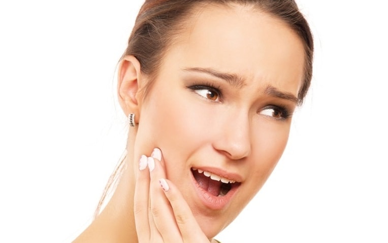
Mechanical damage can be caused by acute trauma as a result of biting the mucous membrane while eating, an epileptic attack, a blow, preparing teeth for crowns (bur, probe, disk), filling teeth, as well as wounding it with a knife, fork, bone, etc.
Acute trauma SOPR can be:
1) open, i.e. with violations of the integrity of the oral mucosa and epithelial cover;
2) closed, i.e. without violating the integrity of the oral mucosa and the epithelial cover.
Open wounds
Occurs more often in practically healthy people from the immediate impact of a traumatic agent and quickly disappear after its elimination.
Depending on the strength and duration of the traumatic factor, the following may occur:
1) excoriation (abrasions) (the layer of the mucous membrane itself is not affected);
2) erosion (surface layers are involved in the process);
Excoriation is a lesion in which the layer of the mucous membrane itself is not affected, expressed pain symptom, but there may not be bleeding, which indicates that the papillary layer is not opened.
Erosion is a superficial damage when the epithelial and papillary layer is involved, which is explained by the appearance of droplets of blood, like “dew”.
Depending on the active factor, the damaging agent, the wound can be:
1) chopped;
2) chopped;
3) torn;
4) bitten.
The clinical manifestations of these wounds depend on the depth of the lesion, type of injury, and vascular involvement. Thus, a bite wound is the most severe, as there is a strong infection. During a bite, up to 170 types of pathogens enter the wound. The very type of wound (laceration) contributes to a significant disruption of microcirculation. Healing is usually progressing secondary intention, through granulation tissue, with the formation of scars, sometimes deforming the skin.
The course of an open wound, regardless of the type of damage, goes through the following stages:
1) the stage of hydration (exudation), which lasts 1–2 days. Patients complain of burning, pain, aggravated by eating and talking. There is pronounced hyperemia and swelling around the lesion. Immediately after an injury, you can apply ice or a cold compress to the wound. Pain is relieved by using painkillers. The wound is washed with antiseptic solutions. Decongestants can be used in cases of extensive lesions (even diuretics). To prevent wound infection, anti-inflammatory drugs are prescribed;
2) stage of dehydration (after 1–3 days). The pain subsides. This stage is characterized by the formation of crusts on skin and a raid on the SOPR. During this period, in addition to anti-inflammatory drugs, enzymes can be prescribed that clean the wound surface of crusts, scales, and plaque; N.B.!!! if the wound heals by secondary intention, then enzymes are contraindicated, since they can melt young, newly formed granulation tissue. Microcirculation is improved by ASA and heparin. Acemin, dibunol improve proliferative regenerative processes;
3) epithelialization stage. Epithelization of acute traumatic lesions occurs quickly, within 1–3 days. When a secondary infection occurs, they long time don't heal. Healing is possible through scarring. Well-proven reparants: vitamins A, E, groups B, C, K, their oil solutions containing O 2, keratoplasty. Promotes regeneration solcoseryl, honsuride, methyl uracil, sodium nucleinate, pentoxyl, actovigen, aloe or colanchoe juice, vinylin, Shestakovsky balm.
Closed wounds
A closed wound is a hematoma - hemorrhage into the tissue surrounding the vessels. The hematoma undergoes changes over several stages, which are called the stages of the hematoma:
1) red hematoma – 1st day. The color of the hematoma is due to hemorrhage of red blood cells into the surrounding tissue. In case of injury, blood vessels rupture, thrombosis, and blood cells leak out. Immediately after the injury, it is good to apply cold and perform cryoapplication. Adrenaline, mezaton, galazalin, ephedrine, sanarin, naphthyzine - topically, especially if the musculoskeletal system is damaged. Ultrasound, laser, UHF, Darsanval currents;
2) blue hematoma – 2-3rd day – explained venous stagnation, change shaped elements. It is good to use FTL, anti-inflammatory therapy, absorbable agents (bodyagu, heparin) during this period;
3) green hematoma – 4-5 days. The color is due to the formation and release of hemasiderin;
author D. N. OrlovLECTURE No. 7. Chronic focal infection of the oral cavity. Diseases of the oral mucosa Chronic infection of the oral cavity has been a subject of increased interest for doctors since ancient times as possible reason many somatic diseases. For the first time the thought that
From the book Dentistry: lecture notes author D. N. Orlov1. Diseases of the oral mucosa Lesions of the oral mucosa are, as a rule, local in nature and can manifest themselves locally and common features(headaches, general weakness, increased body temperature, lack of appetite);
From the book Dentistry: lecture notes author D. N. Orlov1. Acute mechanical trauma Mechanical damage can be caused by acute trauma as a result of biting the mucous membrane while eating, an attack of epilepsy, a blow, preparing teeth for crowns (bur, probe, disk), filling teeth, as well as wounding it
From the book Dentistry: lecture notes author D. N. Orlov2. Chronic mechanical injury (CMT) They are more common than acute ones. Mainly called by the following effective reasons: carious teeth, poor-quality fillings, dentures and their clasps, lack of contact point, tartar, harmful
From the book Dentistry author D. N. Orlov20. Diseases of the oral mucosa Lesions of the oral mucosa are, as a rule, local in nature and can manifest themselves with local and general symptoms (headaches, general weakness, fever, lack of appetite); V
From the book Dentistry author D. N. Orlov25. Acute mechanical trauma of the oral mucosa Mechanical damage can be caused by acute trauma as a result of biting the mucous membrane while eating, an attack of epilepsy, a blow, preparation of teeth for crowns (bur, probe, disk), when
From the book Dentistry author D. N. Orlov26. Chronic mechanical injury (CMT) of the oral mucosa They are more common than acute ones. Caused mainly by the following active reasons: carious teeth, poor-quality fillings, dentures and their clasps, lack of contact
From the book Dentistry author D. N. Orlov28. Chronic chemical injury (CIT) of the oral mucosa Chronic chemical injuries of the mucous membrane have a special manifestation pattern. In some cases they may be in the form allergic reaction delayed type, in others - in the form of intoxication
From the book Cancer: You Have Time author Mikhail Shalnov2. Precancerous diseases of the mucous membrane of the lip, tongue and oral cavity A person comes into contact with the outside world through the oral cavity, accordingly, it is there that inflammatory processes are most likely to develop, which can become the main factors in the development
From the book Homeopathy. Part II. Practical recommendations to the choice of medications by Gerhard KöllerInflammation of the mucous membrane of the mouth and gums Inflammation of the mucous membrane of the mouth and gums occurs in the form of different phases, distinguished by the “pattern” of the mucous membrane. The degree of disruption of the body’s defenses determines the nature of the changes: acute inflammation with
author Evgeniy Vlasovich Borovsky3.1.2. Functions of the oral mucosa The mucous membrane, due to its anatomical and histological features, performs a number of functions: protective, plastic, sensitive, absorptive. Protective function. This function mucous membrane is carried out through a number of mechanisms.
From the book Therapeutic Dentistry. Textbook author Evgeniy Vlasovich Borovsky11.2.1. Mechanical injury This injury can be acute, when the mucous membrane is damaged by a short-term, but significant factor, and chronic - when exposed to a weak irritant for a long time.11.2.1.1. Acute mechanical injuryAcute
From the book Skin and venereal diseases author Oleg Leonidovich IvanovChapter XXVII LESIONS OF THE ORAL MUCOSA AND THE RED BORDER OF THE LIPS Lesions of the oral mucosa occur in many skin diseases and are described in the relevant sections (lichen planus, lupus erythematosus, exudative multiforme
From book Folk remedies in the fight against 100 diseases. Health and longevity author Yu. N. NikolaevInflammation of the oral mucosa 1. Calamus. 1 teaspoon of calamus, finely chopped, infuse with 1.5 cups of boiling water, strain. Rinse your mouth 3 times a day 30 minutes before meals.2. Thick-leaved star anise. Pour 2 tablespoons of chopped rhizomes into 1 glass
From the book Dentistry of Dogs author V.V. Frolov From the book How I cured diseases of the teeth and oral cavity. Unique tips, original techniques author P.V. ArkadyevMost people diligently take care of their teeth, completely forgetting to monitor the health of their gums. Dentists say that the beauty of a smile depends on the condition of the soft tissues in the mouth, so any disease brings serious consequences. The appearance of a dark bruise on the mucous membrane is always alarming and confusing. This may be a consequence of injury or tooth extraction, but in any case it requires careful consideration and full treatment.
A bruise on the gum itself is not separate disease. This is a hemorrhage in soft fabrics mucous membrane due to the destruction of small capillaries. Despite its small size, this problem sometimes provokes serious inflammation in the periosteum and may well cause loss healthy teeth. If it is accompanied by other painful symptoms, requires treatment under the supervision of a dentist.
Most probable reasons formation of a dark spot on the gum in an adult patient:
- The doctor performs manipulations with soft tissues or teeth: removal, filling or cleaning of dental canals, cutting for removal, installation of a prosthesis. The larger the area where such procedures are performed, the more noticeable the bruise on the jaw.
- Problems with blood pressure, which provokes sharp jumps and changes in the circulatory system.
- Congenital diseases associated with blood clotting disorders in the body. In such people, a hematoma on the gum is formed from light biting or.
- Consequences of injury and impact.
In adult patients, a common cause of bruising on the mucous membrane is improper growth of wisdom teeth. As a rule, they come to the surface already mature age when the jaw is fully formed. They do not have enough space in the row, they move to the side and damage the periosteum. In this case, swelling accumulates around the dark spot, and severe redness. The process is accompanied by aching pain, a slight increase in temperature and loss of performance.
Why does a bruise appear on a baby’s gum?
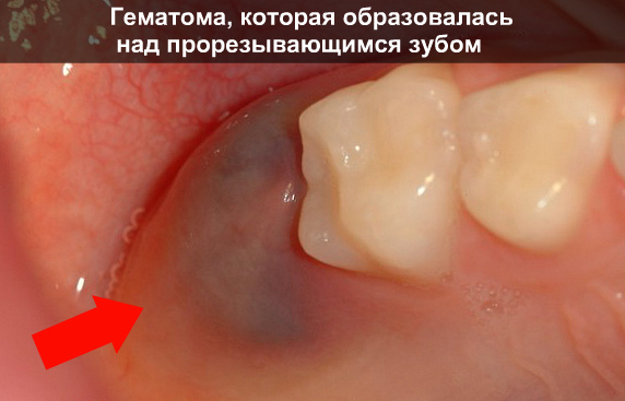
Many parents are perplexed when they discover oral cavity child characteristic bruising. It can be a consequence of injury when active game, chewing on toys or biting on a pacifier. But most often, a small black-blue spot forms in the upper part of the gum.
Dentists warn that teeth sometimes cut through this way. Primary molars and incisors do not have enough sharpness to penetrate thick fabrics periosteum. Damage to the smallest capillaries and slight bleeding into the gum occurs. From the outside, the picture looks unpleasant and worries parents, but in most cases the lump resolves on its own and does not require special treatment. The doctor may recommend light gels with an analgesic so that the baby is not overly restless and can more easily endure the teething process.
Symptoms of bruises in the mouth
Depending on the reason why a hematoma appeared on the gum of an adult, it has different sizes. Dentists divide them into two groups according to the complexity and severity of the consequences:
- Blood pours into the dental pockets and accumulates around the tooth, surrounding it with a dark bluish area. Usually the mucous membrane increases in volume, the patient feels discomfort and cannot completely close the jaw. He finds it difficult to chew and pronounce certain sounds.
- Hemorrhage occurs inside the roots of the teeth, resulting in a rounded swelling affecting the inner surface of the cheek. Outwardly it resembles acute stage: a person complains about aching pain, irritability, slight asymmetry of the face appears. The gums become swollen and there is a feeling of tightness, chills, general weakness and fever.
Gradually with proper treatment All unpleasant symptoms go away, but a spot with a yellowish tint remains at the site of the hematoma. It gradually dissolves and disappears completely. This process may take several weeks.
Methods of treatment for bruises on the gums
Patients mistakenly believe that a bruise should go away without treatment. But a hematoma on the gum after a blow or tooth extraction requires special attention: V open wound Harmful bacteria that are present in the oral cavity easily penetrate. This is fraught with extensive abscesses, fistulas on the mucous membrane and accumulation of suppuration in the roots of the teeth. Therefore, if the temperature rises and discomfort, it is better to seek help.
The dentist may open the gum to allow any accumulated fluid to escape. If necessary, a small drainage is left in the wound to accelerate the outflow of ichor. Through a miniature incision, the doctor washes the damaged area with antiseptics, which eliminates the formation of infection. At home, the patient is recommended to continue therapy:
To heal the incision and speed up the resorption of the bruise, dentists recommend using compresses with healing herbs. After a few days, your gums will become firmer and the dark area will be less sensitive.
Traditional methods of eliminating hematomas on the gums
To improve the health of the mucous membrane, you can use recipes with natural ingredients. Their medicinal properties help to quickly cope with a bruise and restore the beauty of your smile. To prepare natural preparations, plants with wound healing and anti-inflammatory properties are used. Tannins and beneficial compounds promote rapid resorption of hematoma and restore soft periodontal tissue:
- Several calendula inflorescences (fresh or dried) are infused in a glass of boiling water, and the teeth are rinsed 4-5 times a day.
- A decoction of common sage effectively relieves puffiness. It is used warm and try to keep it in the mouth for 5-7 minutes.
- Brewed dry chamomile flowers in combination with St. John's wort will replace the antiseptic and improve the microflora.
- A bruise on the gum is lubricated with fir oil, bay leaf, sea buckthorn or tea tree.
- A paste of aloe or Kalanchoe will speed up wound healing after removing the drainage.
- To reduce pain and swelling, apply an ice cube made with the addition of propolis tincture.
A combination of effective homeopathic recipes and drug treatment will help you quickly get rid of the problem, while avoiding unpleasant complications for the patient.
The reason may be long-term injury mucous membrane with sharp edges of teeth, poorly made or outdated dentures, teeth located outside the arch. Traumatic factors can be overhanging edges of fillings, wire splints or ligatures when splinting jaws, and bad habits. In any case, the traumatic factor has the nature of a long-term impact, which triggers and maintains the mechanism of catarrhal inflammation, which has stages of hyperemia, exudation and proliferation. The severity of each of them depends on the strength and duration of the stimulus.
Exudation can be quite pronounced. Exudate can be serous, serous-purulent and purulent. With purulent exudation, the mucous membrane is subject to superficial destruction, which leads to erosion.
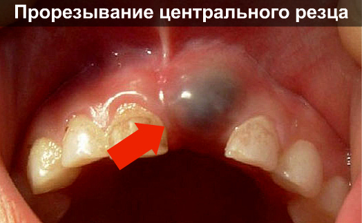
In the absence of treatment, chronic focal purulent inflammation develops. The result of such inflammation is the appearance of a decubital (traumatic) ulcer. Any orthopedic or orthodontic structure can become an inadequate irritant to the oral mucosa. A removable denture transmits chewing pressure to the mucous membrane, delays self-cleaning of the oral cavity, which leads to a change in the established microbial balance. The intermediate part of the bridge has an irritating effect if it touches the mucous membrane of the alveolar edge. Artificial crowns are also an irritant. Even with an ideally made crown, the edges of which are immersed in the gingival groove, it causes injury to the mucous membrane of the gingival margin, which takes the form of a hyperemic dense ridge. Mechanical trauma to the mucous membrane is often associated with teething - the cheek, tongue, lips are bitten, which is determined by the many scars on the oral mucosa; in infants - when feeding from a large nipple.
Clinic
There may be no complaints for a long time, but usually the patient indicates a feeling of awkwardness, discomfort in the oral cavity, minor soreness, and swelling. Depending on the nature of the irritant and the characteristics of the body’s reactivity, changes in the oral mucosa can manifest themselves in the form of catarrhal inflammation, erosion or ulcers. The ulcers are painful, especially when eating and talking. Decubital ulcer is usually single, the mucous membrane around it is swollen, hyperemic, moderately or severely painful. The ulcer has uneven edges and a bottom covered with easily removable fibrinous or necrotic plaque. At the same time, regional The lymph nodes enlarged and painful. Ulcers are most often localized on the mucous membrane of the tongue and cheeks along the line where the teeth meet. With prolonged existence, the edges and base of the ulcer become denser. Its depth can be different, right down to the muscle layer. Inflammation of the mucous membrane of the prosthetic bed can be focal in the form of pinpoint hyperemia or large hyperemic spots and diffuse, often occupying the entire surface of the prosthetic bed. Foci of inflammation usually coincide with areas of incomplete adherence of the prosthesis. Hyperemia of the mucous membrane without violating its integrity is usually observed when using removable dentures for 1-3 years. With longer use, erosions, ulcers, and hyperplastic papillomatous growths occur. Chronic hypertrophic processes occur more often - hypertrophic gingivitis or papillitis, papillomatous growths, prosthetic granuloma, post-traumatic abscess, hyperkeratosis. Prolonged irritation by the edge of the prosthesis can lead to the development of lobular fibroma, which looks like several folds parallel to the edge of the prosthesis. In the presence of poorly manufactured artificial crowns, there is swelling, hyperemia, bleeding of the gingival papillae, deep periodontal pockets with serous or purulent discharge. Clinically, the edematous form of chronic hypertrophic gingivitis or chronic local periodontitis is often determined.
Differential diagnosis
Decubital ulcer must be differentiated from a cancerous ulcer, tuberculous ulcer, chancre, and trophic ulcer. Traumatic erosions and ulcers are differentiated from pemphigus. A traumatic ulcer is painful on palpation and has an inflammatory infiltrate at the base. It is characterized by the presence of an irritating factor, after the elimination of which the ulcer heals in 3-5 days. At cytological examination there are no specific changes. A cancerous ulcer exists for a long time, has dense edges and a bottom, and is characterized by the presence of growths along the edges, resembling in appearance cauliflower, keratinization of the edges. After the appearance of a cancerous ulcer painful sensations weaken. Eliminating the irritant does not lead to healing. Cytological examination reveals atypical cells in the scraping. A tuberculous ulcer is characterized by severe pain and soft, uneven edges. The bottom of the ulcer is granular with yellow spots (Trill grains). Cytological examination reveals giant Langhans cells in scrapings from the surface of the ulcer. A hard chancre has a compaction at the base, its edges are smooth, dense, the bottom is smooth, painless, and flesh-red in color. Regional lymph nodes are enlarged, dense in consistency, mobile and painless. A scraping from an ulcer reveals pale treponema (dark-field microscopy). 3 weeks after the appearance of chancre, it becomes positive reaction Wasserman. Elimination of the traumatic factor, if any, does not affect the course of chancroid, which without treatment can exist for several weeks or even months. A trophic ulcer is characterized by a significant duration of existence, a sluggish course, and the presence of chronic general somatic diseases in the patient. Eliminating the irritating factor does little to promote ulcer healing. In traumatic erosions, unlike pemphigus, Nikolsky's symptom is negative, and there are no acantholytic Tzanck cells in the impression smears. After eliminating the irritant, traumatic ulcers and erosions heal quickly.
Treatment
First of all, it is necessary to eliminate or weaken the effect of the irritating factor. It is prohibited to wear defective removable dentures; rational prosthetics are recommended. It is necessary to replace low-quality fixed structures and fillings. Sanitation of the oral cavity and repeated professional hygiene. In case of severe pain, anesthesia is carried out with any warm anesthetic: 0.5-1% lidocaine solution, 0.5-1% novocaine solution, 2-4% pyromecaine solution, in the form of applications, irrigations, baths or rinses.
If there is necrotic or fibrinous plaque on the surface of an erosion or ulcer, it is recommended to apply proteolytic enzymes for 8-10 minutes, after which the necrotic tissue or fibrinous plaque is removed mechanically, and the ulcer or erosion is treated with antiseptics (0.02% furatsilin solution, 0.5 % solution hydrogen peroxide, 0.5-1% etonium solution, 0.5-1% di-mexide solution). Applications of foam aerosols, methyluracil or solcoseryl ointment are applied, and from the moment epithelization begins, applications of keratoplastics are applied for 15-20 minutes 3-4 times a day (sea buckthorn oil, rose hips, vinylin, Unna or KF cream, vitamin A or E in oil) .
Treatment of lobular fibroma consists of correction of the orthopedic structure, if possible, or rational prosthetics and applications of keratoplastics for 15-20 minutes 3-4 times a day are immediately recommended. If necessary and indicated, the lobular fibroma is excised and a new prosthesis is made.
Treatment of traumatic injuries due to habitual biting of the mucous membrane comes down to eliminating all irritating factors and timely sanitation of the oral cavity, as well as rational prosthetics and monitoring the condition of prostheses. Often, cheek biting, as a bad habit, leads to the development of soft leukoplakia, about which we'll talk below.
The oral mucosa is constantly exposed to various damaging factors. Thanks to its protective function the mucous membrane does not change under impacts not exceeding a certain threshold. When exposed to suprathreshold stimuli, certain changes occur on the mucous membrane, which are classified as traumatic lesions.
Epidemiology
The frequency of occurrence of certain forms of traumatic lesions of the oral mucosa varies depending on the form of the disease.Classification
Traumatic lesions are divided:- by causal influencing factors:
- chemical;
- physical;
- with the flow:
- chronic; - traumatic erythema;
- bubbles;
— erosion;
- ulcer;
- cracked lips;
- keratosis;
- cheilitis;
- hemorrhagic changes (with radiation damage);
— necrosis (with chemical damage);
— leukoplakia: leukoplakia of Tappeiner smokers; flat leukoplakia; verrucous leukoplakia; erosive leukoplakia.
Etiology and pathogenesis
For acute mechanical injury the mucous membrane is affected by a short-term but significant factor. With chronic mechanical injury, the stimulus is weak, but the impact is longer. Traumatic factors can be sharp edges of teeth, bridges, removable dentures, tartar deposits, orthodontic appliances, and bad habits.Chemical injury
Damage occurs upon contact with the mucous membrane chemical substances- concentrated solutions of acids, alkalis, some drugs.Physical trauma
Acute physical injuries occur when thermal burns, exposure to electric current, high doses of ionizing radiation. Chronic physical injury can be caused by meteorological conditions, prolonged exposure to low doses of ionizing radiation, as well as galvanic currents arising in the oral cavity in the presence of dissimilar metals.Gastrointestinal diseases, diabetes, anemia, deficiency or metabolic disorder of vitamin A create the background for the development of leukoplakia. Leading value have local irritants ( tobacco smoke, galvanism, eating very hot food, sharp edges of teeth, low-quality dentures, etc.).
Clinical signs and symptoms
Clinical manifestations of traumatic injuries vary widely depending on the strength of the damaging agent, local conditions, the state of the microbiocenosis, and the general reactivity of the body. With mechanical trauma, changes in the mucous membrane can manifest as catarrhal inflammation, disruption of the integrity of the epithelium, hyperplastic processes, and hyperkeratinization.Chemical injury
In case of burns with acids, coagulation necrosis occurs, in case of burns with alkalis - colliquation necrosis. With chronic chemical trauma, chronic catarrhal inflammation, ulcerative necrotizing gingivostomatitis, keratosis, and leukoplakia are observed.Physical trauma
With acute thermal damage, blisters, erosions, and ulcers appear. Acute electric shock causes long-lasting ulcers. Clinical picture radiation stomatitis consists mainly of hemorrhagic syndrome and ulcerative-necrotic process. With a galvanic current exceeding 10 μA, paresthesia of the mucous membrane, chronic inflammatory processes, as well as complications of existing pathological processes (lichen planus, leukoplakia).Leukoplakia
The clinical picture depends on the form of leukoplakia, the factors that caused it and the location.Flat shape: limited, non-raising areas of keratinization of a grayish-white color without compaction at the base. Areas of flat leukoplakia look like a lapis burn or tissue paper pasted on and are not removed by scraping.
Verrucous form: limited white plaques with an uneven surface (plaque form) or dense warty growths (warty form).
Erosive form: against the background of foci of hyperkeratosis, erosions or cracks are detected. It is a complication of simple or verrucous leukoplakia.
Leukoplakia of smokers: keratinization hard palate, as well as the back of the tongue. After quitting smoking it quickly regresses.
Diagnosis is based on medical history and identification of the irritant. Elimination of the traumatic factor serves differential diagnostic purposes.Additional research methods: luminescent (gray-yellow glow of the lesion in leukoplakia), cytological (to detect atypical cells). If necessary, consultation with a neuropsychiatrist is recommended.
Differential diagnosis
It is necessary to distinguish a traumatic ulcer from a cancerous, tuberculous, syphilitic ulcer, chronic ulcerative-necrotic Vincent gingivostomatitis, trophic ulcer. Leukoplakia is differentiated from lichen planus, lupus erythematosus, candidiasis, soft leukoplakia, secondary syphilis, opacification of the epithelium during its regeneration, and Bowen's disease. For traumatic lesions of the oral mucosa, the basis therapeutic measures is to eliminate the traumatic factor. Sanitation of the oral cavity, avoidance of smoking, consumption of hot food, alcohol, and galvanism are recommended; rational dental prosthetics is indicated. If necessary, in order to get rid of bad habit biting the mucous membrane, consultation with a neuropsychiatrist is recommended.An increase in the size of the focus of flat leukoplakia, the ineffectiveness of conservative measures for the erosive form, as well as the verrucous form of the disease are indications for surgical treatment. Cryodestruction, electrocoagulation, excision of the lesion using laser scalpel. For topical anesthesia to anesthetize the mucous membrane before eating, medicinal treatment of lesions, local anesthetics are used:
Benzocaine/glycerin topically 5/20 g before each meal until clinical improvement or
Lidocaine, 2.5-5% ointment or 10% aerosol, topically before each meal, until clinical improvement.
For pain relief before eating in a benzocaine solution, you can use olive or peach oil instead of glycerin. Antiseptics and antimicrobial drugs are used to treat the oral cavity and damaged mucous membranes and prevent their infection.
Treatment is carried out with loose cotton swabs soaked in a warm antiseptic solution; oral baths are also used:
Hydrogen peroxide, 1% solution, topically 1-2 times or
Potassium permanganate, 0.02% solution, topically 1-2 times or
Chlorhexiddine, 0.06% solution, topically 1-2 times or
Ethacridine, 0.05% solution, topically 1-2 times.
To clean the surface of erosions and ulcers, proteolytic enzymes are used, which are applied to the affected element:
Trypsin 5 mg (in isotonic sodium chloride solution) locally 1-2 times a day or
Chymotrypsin 5 mg (in isotonic sodium chloride solution) locally 1-2 times a day.
If it is necessary to correct the psycho-emotional sphere, sedative therapy is recommended:
Valerian rhizome extract orally 1 tablet 1-2 times a day, long-term or
Glycine sublingually 0.1 g 2-3 times a day, long-term.
It is also possible to use other drugs plant origin. In more severe cases, tranquilizers are used (after consultation with a neuropsychiatrist):
Diazepam orally 5-15 mg 1-2 times a day, 4 weeks or
Medazepam orally 10 mg 2-3 times a day, 4 weeks or
Midazolam orally before bedtime 7.5-15 mg once a day, 4 weeks or
Nitrazepam orally 30-40 minutes before bedtime 5-10 mg 1 time per day, 4 weeks.
Medazepam is prescribed as a daytime tranquilizer, midazolam and nitrazepam as hypnotics. To speed up the healing of affected areas, drugs that stimulate regeneration processes and vitamins are used:
Sea buckthorn oil applied topically to the cleaned area of the affected mucous membrane 1-3 times a day, until clinical improvement or
Solcoseryl, ointment or dental adhesive paste, topically on the cleaned area of the affected mucous membrane 1-3 times a day, until clinical improvement or
Rosehip oil applied topically to the cleaned area of the affected mucous membrane 1-3 times a day, until clinical improvement
+
Retinol, solution, topically on the affected areas 5-6 times a day, until clinical improvement (used as an anti-inflammatory, immunostimulating agent that improves tissue trophism)
+
Vitamin E, solution, topically on the affected areas 5-6 times a day, until clinical improvement (used as an active antioxidant to stimulate protein synthesis and reduce capillary permeability)
+
Ascorbic acid orally 50-100 mg 3-5 times a day or 5% solution IM 1 ml 1 time a day, 20-40 days (used to regulate redox processes, stimulate tissue regeneration, activate phagocytosis and antibody synthesis)
+
Calcium pantothenate orally 0.1 g 2-4 r/day or 5% solution locally in the form of applications for long-term non-healing erosions 2-4 r/day or 10% solution IM 2 ml 1-2 r/ days, 20-40 days (used to normalize fatty acid metabolism, stimulate the formation of acetylcholine, steroid hormones, and utilize amino acid deamination products)
+
Rutoside orally 0.02-0.05 g 3 times a day, 20-40 days (reduces vascular permeability, protects ascorbic acid from oxidation and together with it reduces the activity of hyaluronidase)
+
Cyanocobalamin orally 0.00005 g 1 time / day, 20-40 days
+
Folic acid orally 0.0008 g 1 time / day, 20-40 days (cyanocobalamin and folic acid used to activate the processes of hematopoiesis and maturation of red blood cells, tissue regeneration)
+
Riboflavin orally 0.005-0.01 g 1 time / day, 20-40 days (prescribed to regulate tissue respiration, metabolic processes)
+
Nicotinic acid orally after meals 0.025-0.05 g 3 times / day, 20-40 days or 1% solution IV, IM or under the lesion 1 ml 1 time / day, 10-15 days (used in order to normalize metabolic processes and peripheral blood supply).
Depending on the clinical situation, vitamins can be prescribed in various combinations. As an anti-inflammatory, decongestant, analgesic, wound-healing and immunostimulating agent, you can also use the homeopathic remedy Traumeel S (orally, sublingually, intramuscularly or locally).
Evaluation of treatment effectiveness
Restoring the integrity of the epithelium, the absence inflammatory reaction are evidence successful treatment traumatic lesions of the oral mucosa. The course of treatment should be 10-14 days. In the absence of treatment effect, it is necessary to exclude oncological diseases. Treatment of leukoplakia is considered effective in the transition of verrucous and erosive forms to flat, and in the future - complete elimination of foci of hyperkeratosis.Errors and unreasonable assignments
The main mistake in the treatment of chronic traumatic lesions of the oral mucosa is the prescription of drugs exclusively for self-use patient. Professional medicinal treatment of lesions is mandatory (if the integrity of the epithelium is damaged, it should be carried out daily). The use of irritating and cauterizing agents for leukoplakia is contraindicated.Forecast
With a long course, malignancy of a traumatic ulcer is possible. All forms of leukoplakia can become malignant, the most dangerous erosive form. Prolonged course of chronic radiation sickness leads to radiation periodontitis. Post-radiation stomatitis is an optional precancer. Such patients should be under constant medical supervision.G.M. Barer, E.V. Zoryan
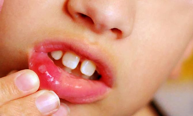
The lining of the oral cavity is the most resistant human mucosa to irritants and pathogenic microorganisms. Therefore she is rarely exposed external influence. A blood blister most often forms as a result of injury. Less commonly, the cause is dental diseases or systemic pathologies.
Bubble on inside cheeks - this is a hematoma, or bruise. It is a small, from a few millimeters to a couple of centimeters, rounded formation. The internal exudate is serous or bloody, depending on the extent of the injury. In the first case, the blister is gray-white, in the second it is red, with a possible bluish tint.
Blood blisters appear due to injuries to the mucous membrane. Their formation is a natural reaction immune system to the stimulus. It consists in:
- Activations defense mechanisms body in response to a stimulus. Agranulocytes arrive at the site of the lesion: leukocytes and monocytes. The latter, when leaving the blood into the tissues, are converted into macrophages. These cells capture the pathogen, neutralize it, and then die.
- The death of agranulocytes leads to their release to the site of injury. This process is a signal of injury to the body. After which it begins to release histamine, bradykinin and serotonin into the affected area. They sharply increase the permeability of blood vessels.
- A spasm occurs at the site of injury. The blood flow becomes difficult, after which the vessels relax and the blood accumulated during the narrowing enters the injured area. It moves quickly and under pressure. As a result, the upper layer of the epithelium peels off and a blister filled with bloody contents forms.
Blood blisters appear due to injuries to the mucous membrane.
Important! danger red or white growth does not carry. It is virtually painless and heals within a week. The only discomfort is associated with inconvenience when chewing and talking.
Causes
The most common reason Why blood blisters appeared in the mouth is an accidental injury to the mucous membrane. There are three possible options lesions:
Mechanical
A blood blister is formed when accidentally biting while talking or eating. The mucous membrane can also be injured:
- hard foods: candies, crackers, bones;
- broken, chipped teeth or incorrectly made dental structures: braces, bridges, crowns, dentures;
- during hygiene procedures– tissues are damaged due to careless movements while brushing your teeth with a brush or toothpick.
In addition, you can bite your cheek while epileptic seizure, in a dream or with strong excitement. In these cases, the patient may not remember the moment the injury occurred.
A blister may appear after biting with your teeth.
Important! Less commonly, a bloody blister can form due to dental surgery. It is associated with careless actions of the doctor during sanitation of the oral cavity.
Thermal
A blister on the cheek can form due to burns from hot drinks, dishes, inhalation of steam or accidental contact with heated cutlery. In this case, the appearance of a bubble is accompanied by a burning sensation, swelling, redness and slight pain.
Chemical
The cause is tissue damage by aggressive chemical elements: In case of accidental ingestion or inhalation of vapors at home or at work, bubbles may appear. As with thermal injury, mucosal hyperemia and painful sensations are observed.
Important! Provoking factors for the appearance of blisters in the oral cavity include smoking, abuse of strong alcohol, and hypovitaminosis. It is believed that under the influence of harmful elements and a lack of vitamins, the walls of blood vessels become thinner. This provokes hemorrhage and the formation of hematomas.
Traumatic causes are typical for single bloody or serous formations. If the bubbles appear regularly, there are many of them, they are localized not only on the cheek, but also on the tongue, gums, lips, and are accompanied by other symptoms (plaque, itching, unpleasant smell) – this indicates diseases of the oral cavity or systemic pathologies. Among these factors are:

Treatment
Usually a blood blister is not required specific treatment. Since it is a natural response of the immune system to external stimuli, the formation goes away on its own within a few days.
Important! If the resulting blister did not appear due to injury, often recurs, or multiple growths are noted, a doctor’s consultation is required to rule out possible pathologies.
In some cases, therapy for hematoma is indicated. It is required when large size bubble, soreness and discomfort. When treating, the dentist takes into account the following factors:
- size of education;
- time and factor of appearance;
- location of the bubble: on the cheek, tongue, lip, gums;
- whether there are other blisters or sores.
If the bubble does not go away, contact your dentist.
Treatment of a bloody ball consists of a puncture, ensuring the outflow of accumulated fluid and antiseptic treatment. Rarely, surgical excision of tissue is required to remove the lesion.
When blisters appear due to chipped teeth or incorrect dental work, the defects must be corrected. Otherwise, the cheek will be constantly injured.
Important! If the doctor suspects that the bubbles have formed not due to injuries, but as a consequence of systemic pathologies, the patient will be prescribed comprehensive examination. Further therapy will be based on test results.
According to indications, multivitamin complexes with a high content of vitamins C, K, E, A, and group B can be prescribed. This will strengthen the walls of blood vessels and prevent hemorrhages.
After surgical intervention the patient is recommended:

What can and cannot be done if a blister appears on the cheek?
The formation of a blood globule is always a cause for concern. However, there is no need to panic. First of all, it is necessary to establish the reason why it could appear: whether there were injuries, whether hot or irritating food was consumed. Further actions aimed at relieving inflammation and disinfection:
- The mouth is treated with antiseptic drugs.
- Rinsing with a solution of baking soda and salt will help relieve inflammation.
- Provoking factors are excluded: smoking, alcohol, consumption of salty, sour, spicy, pickled foods.
If after a few days the ball does not shrink and signs of healing are not visible, you should consult a doctor: dentist or therapist.
Bloody blisters in the mouth most often appear as a result of injuries: biting, burns, chemical injuries. Less common factors are diseases of the oral cavity and systemic pathologies. Usually, special treatment not required. If the formation interferes, the dentist pierces it and prescribes antiseptic treatment.

 Echo HSG tubal patency")


